Explore
 Validate
 Learn
Learn Western blot
 Flow cytometry
Antibody data
- Antibody Data
- Antigen structure
- References [4]
- Comments [0]
- Validations
- Western blot [2]
- Immunocytochemistry [1]
- Immunohistochemistry [1]
Submit
Validation data
Reference
Comment
Report error
- Product number
- GTX108152 - Provider product page

- Provider
- GeneTex
- Proper citation
- GeneTex Cat#GTX108152, RRID:AB_2037202
- Product name
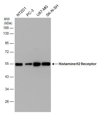
- Histamine H2 Receptor antibody [N1], N-term
- Antibody type
- Polyclonal
- Reactivity
- Human, Rat
- Host
- Rabbit
Submitted references Suppression of osteogenic differentiation in mesenchymal stem cells from patients with ossification of the posterior longitudinal ligament by a histamine-2-receptor antagonist.
Involvement of the H1 Histamine Receptor, p38 MAP Kinase, Myosin Light Chains Kinase, and Rho/ROCK in Histamine-Induced Endothelial Barrier Dysfunction.
Involvement of H1 and H2 receptors and soluble guanylate cyclase in histamine-induced relaxation of rat mesenteric collecting lymphatics.
Histamine receptor 2 modifies dendritic cell responses to microbial ligands.
Liu X, Kumagai G, Wada K, Tanaka T, Fujita T, Sasaki A, Furukawa KI, Ishibashi Y
European journal of pharmacology 2017 Sep 5;810:156-162
European journal of pharmacology 2017 Sep 5;810:156-162
Involvement of the H1 Histamine Receptor, p38 MAP Kinase, Myosin Light Chains Kinase, and Rho/ROCK in Histamine-Induced Endothelial Barrier Dysfunction.
Adderley SP, Zhang XE, Breslin JW
Microcirculation (New York, N.Y. : 1994) 2015 May;22(4):237-48
Microcirculation (New York, N.Y. : 1994) 2015 May;22(4):237-48
Involvement of H1 and H2 receptors and soluble guanylate cyclase in histamine-induced relaxation of rat mesenteric collecting lymphatics.
Kurtz KH, Moor AN, Souza-Smith FM, Breslin JW
Microcirculation (New York, N.Y. : 1994) 2014 Oct;21(7):593-605
Microcirculation (New York, N.Y. : 1994) 2014 Oct;21(7):593-605
Histamine receptor 2 modifies dendritic cell responses to microbial ligands.
Frei R, Ferstl R, Konieczna P, Ziegler M, Simon T, Rugeles TM, Mailand S, Watanabe T, Lauener R, Akdis CA, O'Mahony L
The Journal of allergy and clinical immunology 2013 Jul;132(1):194-204
The Journal of allergy and clinical immunology 2013 Jul;132(1):194-204
No comments: Submit comment
Supportive validation
- Submitted by
- GeneTex (provider)
- Main image
- Experimental details
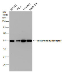
- Histamine H2 Receptor antibody [N1], N-term detects HRH2 protein by Western blot analysis.A. 30 µg U87-MG whole cell lysate/extractB. 30 µg SK-N-SH whole cell lysate/extract10 % SDS-PAGEHistamine H2 Receptor antibody [N1], N-term (GTX108152) dilution: 1:1000
- Validation comment
- WB
- Submitted by
- GeneTex (provider)
- Main image
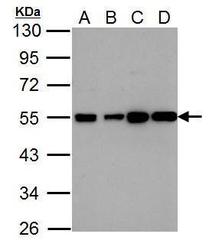
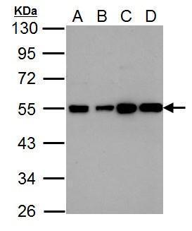
- Experimental details
- Various whole cell extracts (30 ?g) were separated by 10% SDS-PAGE, and the membrane was blotted with Histamine H2 Receptor antibody [N1], N-term (GTX108152) diluted at 1:1000.
Supportive validation
- Submitted by
- GeneTex (provider)
- Main image

- Experimental details
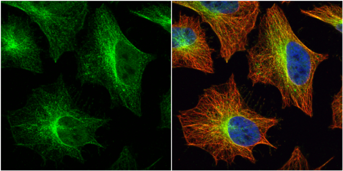
- Histamine H2 Receptor antibody [N1], N-term detects Histamine H2 Receptor protein at cytoplasm by immunofluorescent analysis.Sample: HeLa cells were fixed in 4% paraformaldehyde at RT for 15 min.Green: Histamine H2 Receptor protein stained by Histamine H2 Receptor antibody [N1], N-term (GTX108152) diluted at 1:500.Red: alpha Tubulin, a cytoskeleton marker, stained by alpha Tubulin antibody [GT114] (GTX628802) diluted at 1:1000.Blue: Hoechst 33342 staining.
Supportive validation
- Submitted by
- GeneTex (provider)
- Main image

- Experimental details
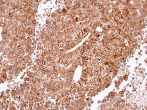
- Immunohistochemical analysis of paraffin-embedded H520 xenograft, using Histamine H2 Receptor(GTX108152) antibody at 1:100 dilution.