Explore
 Validate
 Learn
Learn Western blot
 Immunocytochemistry
Antibody data
- Antibody Data
- Antigen structure
- References [3]
- Comments [0]
- Validations
- Immunocytochemistry [7]
- Other assay [5]
Submit
Validation data
Reference
Comment
Report error
- Product number
- PA5-19474 - Provider product page

- Provider
- Invitrogen Antibodies
- Product name
- PADI2 Polyclonal Antibody
- Antibody type
- Polyclonal
- Antigen
- Synthetic peptide
- Description
- For Western Blot, this antibody has non-specific bands at 34 kDa. Heat mediated antigen retrieval recommended prior to tissue staining.
- Reactivity
- Human, Rat
- Host
- Rabbit
- Isotype
- IgG
- Vial size
- 100 μg
- Concentration
- 0.9 mg/mL
- Storage
- Store at 4°C short term. For long term storage, store at -20°C, avoiding freeze/thaw cycles.
Submitted references Activation of Peptidylarginine Deiminase in the Salivary Glands of Balb/c Mice Drives the Citrullination of Ro and La Ribonucleoproteins.
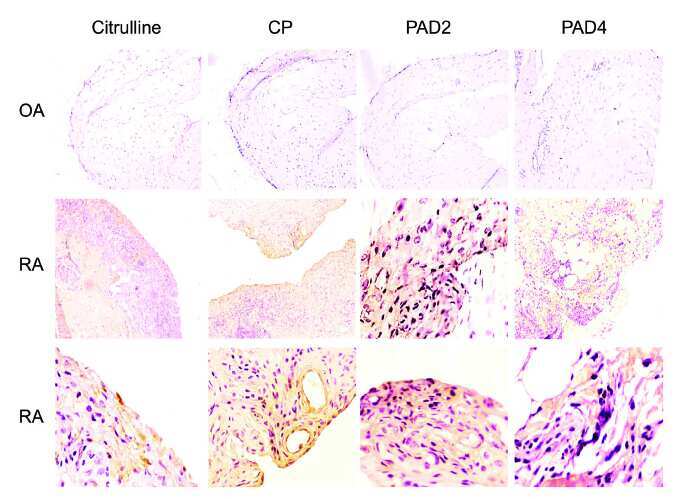
Potential protein targets of the peptidylarginine deiminase 2 and peptidylarginine deiminase 4 enzymes in rheumatoid synovial tissue and its possible meaning.
Posttranslational Protein Modification in the Salivary Glands of Sjögren's Syndrome Patients.
Rodríguez-Rodríguez M, Herrera-Esparza R, Bollain Y Goytia JJ, Pérez-Pérez ME, Pacheco-Tovar D, Murillo-Vázquez J, Pacheco-Tovar G, Avalos-Díaz E
Journal of immunology research 2017;2017:8959687
Journal of immunology research 2017;2017:8959687
Potential protein targets of the peptidylarginine deiminase 2 and peptidylarginine deiminase 4 enzymes in rheumatoid synovial tissue and its possible meaning.
Badillo-Soto MA, Rodríguez-Rodríguez M, Pérez-Pérez ME, Daza-Benitez L, Bollain-Y-Goytia JJ, Carrillo-Jiménez MA, Avalos-Díaz E, Herrera-Esparza R
European journal of rheumatology 2016 Jun;3(2):44-49
European journal of rheumatology 2016 Jun;3(2):44-49
Posttranslational Protein Modification in the Salivary Glands of Sjögren's Syndrome Patients.
Herrera-Esparza R, Rodríguez-Rodríguez M, Pérez-Pérez ME, Badillo-Soto MA, Torres-Del-Muro F, Bollain-Y-Goytia JJ, Pacheco-Tovar D, Avalos-Díaz E
Autoimmune diseases 2013;2013:548064
Autoimmune diseases 2013;2013:548064
No comments: Submit comment
Supportive validation
- Submitted by
- Invitrogen Antibodies (provider)
- Main image

- Experimental details
- Immunofluorescent staining of HEK293 cells using Product # PA5-19474, anti-PADI2/PAD2 antibody. The cells were fixed with PFA (4%)for 10 minutes, permabilised PBS-T for 20 minutes and exposed to the primary antibody at a concentration of 5 µg/mL for 1 hour at room temp. A solution of BSA (1%), normal goat serum (10%) and glycine (0.3 M) was used to quench autofluorescence and block non-specific protein-protein interactions. The secondary antibody was a 448 fluorescence conjugated Goat anti-rabbit IgG (green) at a dilution of 1:1000. A WGA- 594 fluorescent conjugated stain was used to label plasma membranes (red) and the nuclei stain was DAPI (blue).

- Submitted by
- Invitrogen Antibodies (provider)
- Main image

- Experimental details
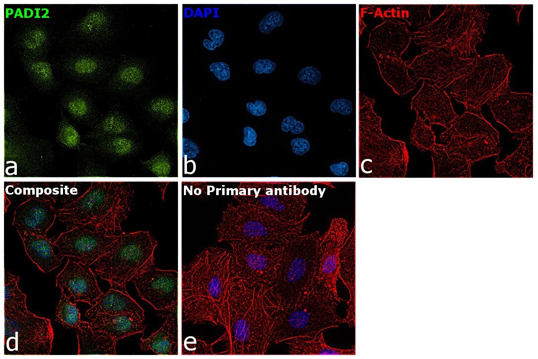
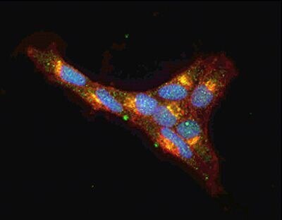
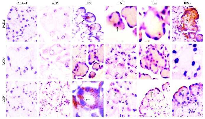
- Immunofluorescence analysis of PADI2 was performed using 70% confluent log phase A-549 cells. The cells were fixed with 4% paraformaldehyde for 10 minutes, permeabilized with 0.1% Triton™ X-100 for 15 minutes, and blocked with 2% BSA for 1 hour at room temperature. The cells were labeled with PADI2 Polyclonal Antibody (Product # PA5-19474) at 1:100 dilution in 0.1% BSA and incubated overnight at 4 degree and then labeled with Goat anti-Rabbit IgG (H+L) Superclonal™ Recombinant Secondary Antibody, Alexa Fluor® 488 conjugate (Product # A27034) at a dilution of 1:2000 for 45 minutes at room temperature (Panel a: green). Nuclei (Panel b: blue) were stained with ProLong™ Diamond Antifade Mountant with DAPI (Product # P36962). F-actin (Panel c: red) was stained with Rhodamine Phalloidin (Product # R415, 1:300). Panel d represents the composite image showing nuclear localization. Panel e represents control cells with no primary antibody to assess background. The images were captured at 60X magnification.
- Submitted by
- Invitrogen Antibodies (provider)
- Main image

- Experimental details
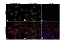
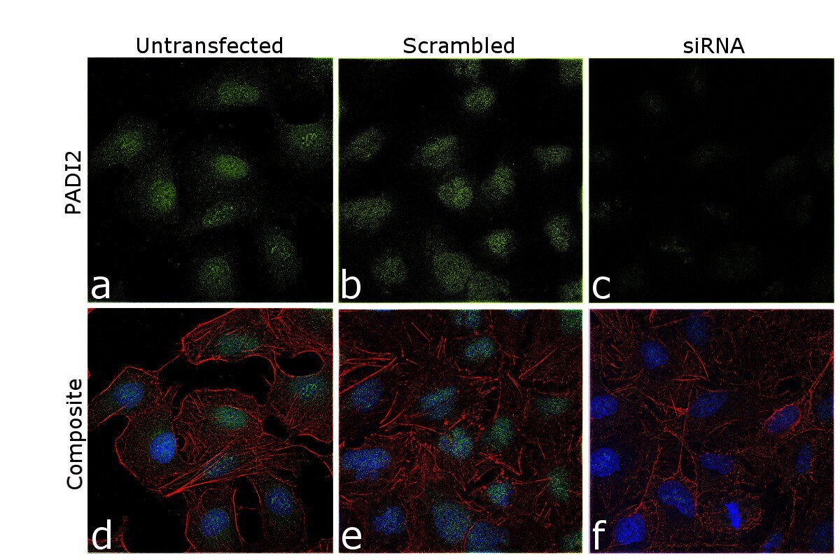
- Knockdown of PADI2 was achieved by transfecting A-549 cells with PADI2 specific siRNA (Silencer® select Product # s22188, Product # s22187). Immunofluorescence analysis was performed on A549 cells (untransfected, panel a,d), transfected with non-specific scrambled siRNA (panels b,e) and transfected with PADI2 specific siRNA (panel c,f). Cells were fixed, permeabilized, and labelled with PADI2 Polyclonal Antibody (Product # PA5-19474, 1:100 dilution), followed by Goat anti-Rabbit IgG (H+L) Superclonal™ Recombinant Secondary Antibody, Alexa Fluor® 488 conjugate (Product # A27034, 1:2000). Nuclei (blue) were stained using ProLong™ Diamond Antifade Mountant with DAPI (Product # P36962), and Rhodamine Phalloidin (Product # R415, 1:300) was used for cytoskeletal F-actin (red) staining. Reduction of specific signal was observed upon siRNA mediated knockdown (panel c,f) confirming specificity of the antibody to PADI2 (green). The images were captured at 60X magnification.

- Submitted by
- Invitrogen Antibodies (provider)
- Main image
- Experimental details
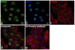
- Immunofluorescence analysis of PADI2 was performed using 70% confluent log phase A-549 cells. The cells were fixed with 4% paraformaldehyde for 10 minutes, permeabilized with 0.1% Triton™ X-100 for 15 minutes, and blocked with 2% BSA for 1 hour at room temperature. The cells were labeled with PADI2 Polyclonal Antibody (Product # PA5-19474) at 1:100 dilution in 0.1% BSA and incubated overnight at 4 degree and then labeled with Goat anti-Rabbit IgG (Heavy Chain) Superclonal™ Recombinant Secondary Antibody, Alexa Fluor® 488 conjugate (Product # A27034) at a dilution of 1:2000 for 45 minutes at room temperature (Panel a: green). Nuclei (Panel b: blue) were stained with ProLong™ Diamond Antifade Mountant with DAPI (Product # P36962). F-actin (Panel c: red) was stained with Rhodamine Phalloidin (Product # R415, 1:300). Panel d represents the composite image showing nuclear localization. Panel e represents control cells with no primary antibody to assess background. The images were captured at 60X magnification.

- Submitted by
- Invitrogen Antibodies (provider)
- Main image
- Experimental details
- Knockdown of PADI2 was achieved by transfecting A-549 cells with PADI2 specific siRNA (Silencer® select Product # s22188, Product # s22187). Immunofluorescence analysis was performed on A549 cells (untransfected, panel a,d), transfected with non-specific scrambled siRNA (panels b,e) and transfected with PADI2 specific siRNA (panel c,f). Cells were fixed, permeabilized, and labelled with PADI2 Polyclonal Antibody (Product # PA5-19474, 1:100 dilution), followed by Goat anti-Rabbit IgG (Heavy Chain) Superclonal™ Recombinant Secondary Antibody, Alexa Fluor® 488 conjugate (Product # A27034, 1:2000). Nuclei (blue) were stained using ProLong™ Diamond Antifade Mountant with DAPI (Product # P36962), and Rhodamine Phalloidin (Product # R415, 1:300) was used for cytoskeletal F-actin (red) staining. Reduction of specific signal was observed upon siRNA mediated knockdown (panel c,f) confirming specificity of the antibody to PADI2 (green). The images were captured at 60X magnification.
- Submitted by
- Invitrogen Antibodies (provider)
- Main image

- Experimental details
- Immunofluorescent staining of HEK293 cells using Product # PA5-19474, anti-PADI2/PAD2 antibody. The cells were fixed with PFA (4%)for 10 minutes, permabilised PBS-T for 20 minutes and exposed to the primary antibody at a concentration of 5 µg/mL for 1 hour at room temp. A solution of BSA (1%), normal goat serum (10%) and glycine (0.3 M) was used to quench autofluorescence and block non-specific protein-protein interactions. The secondary antibody was a 448 fluorescence conjugated Goat anti-rabbit IgG (green) at a dilution of 1:1000. A WGA- 594 fluorescent conjugated stain was used to label plasma membranes (red) and the nuclei stain was DAPI (blue).
Supportive validation
- Submitted by
- Invitrogen Antibodies (provider)
- Main image
- Experimental details
- NULL
- Submitted by
- Invitrogen Antibodies (provider)
- Main image
- Experimental details
- NULL
- Submitted by
- Invitrogen Antibodies (provider)
- Main image
- Experimental details
- NULL
- Submitted by
- Invitrogen Antibodies (provider)
- Main image

- Experimental details
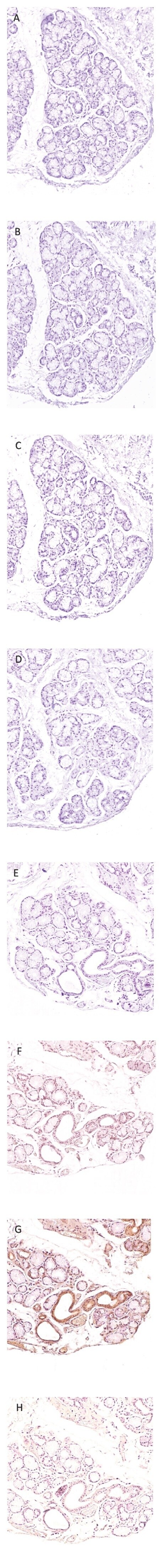
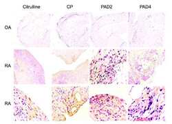
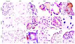
- Figure 4 Immunohistochemistry of minor salivary glands from Sjogren patients; the superior panel shows an overview of immunoreagent distribution at 10x magnification; the inferior panel showed a close up at 40x magnification. (A) and (E). Incubated with PBS. (B) and (F). Treated with anti-CCP. (C) and (G). Anti-PAD2. (D) and (H). Anticitrulline. Note the correlation between CCP, PAD2, and citrulline expression in a duct.

- Submitted by
- Invitrogen Antibodies (provider)
- Main image

- Experimental details
- Figure 2 Immunohistochemistry of minor salivary glands. The superior panel corresponds to normal controls, and the inferior panel belongs to one representative biopsy of a Sjogren patient. (A) and (E). Incubated with PBS. (B) and (F). Treated with anti-CCP. (C) and (G). Anti-PAD2. (D) and (H). Anticitrulline. Observe the immunoreagents positivity at ducts and acini in (F), (G), and (H).