Explore
 Validate
 Learn
Learn Western blot
Antibody data
- Antibody Data
- Antigen structure
- References [1]
- Comments [0]
- Validations
- Western blot [4]
- Immunocytochemistry [1]
- Immunohistochemistry [3]
- Other assay [1]
Submit
Validation data
Reference
Comment
Report error
- Product number
- PA5-77566 - Provider product page

- Provider
- Invitrogen Antibodies
- Product name
- Orexin Receptor 1 Polyclonal Antibody
- Antibody type
- Polyclonal
- Antigen
- Synthetic peptide
- Description
- For reconstitution, we recommend adding 100 µL distilled water to a final antibody concentration of about 1 mg/mL. To use this carrier-free antibody for conjugation experiments, we strongly recommend performing another round of desalting. (Zeba Spin Desalting Columns, 7KMWCO, 0.5 mL, Product # 89882)
- Reactivity
- Human, Mouse, Rat
- Host
- Rabbit
- Isotype
- IgG
- Vial size
- 50 µL
- Concentration
- 0.8 mg/mL
- Storage
- -20°C
Submitted references Orexin A alleviates neuroinflammation via OXR2/CaMKKβ/AMPK signaling pathway after ICH in mice.
Li T, Xu W, Ouyang J, Lu X, Sherchan P, Lenahan C, Irio G, Zhang JH, Zhao J, Zhang Y, Tang J
Journal of neuroinflammation 2020 Jun 15;17(1):187
Journal of neuroinflammation 2020 Jun 15;17(1):187
No comments: Submit comment
Supportive validation
- Submitted by
- Invitrogen Antibodies (provider)
- Main image

- Experimental details
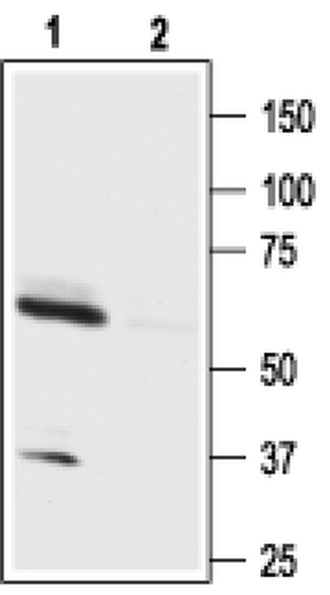
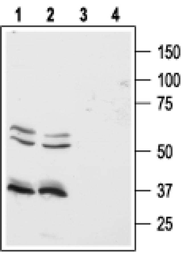
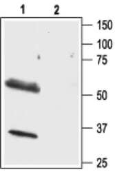
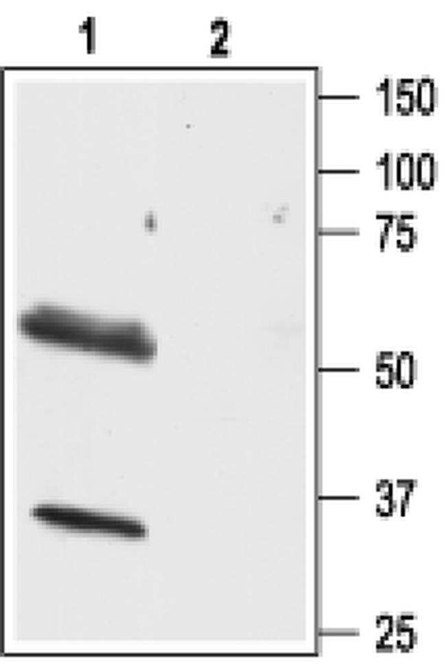
- Western blot analysis of human Colo-205 (lanes 1 and 3) and HT-29 (lanes 2 and 4) colon cancer cell lines with Orexin Receptor 1 polyclonal antibody (Product # PA5-77566) using a dilution of 1:500.

- Submitted by
- Invitrogen Antibodies (provider)
- Main image

- Experimental details
- Western blot analysis of human Colo-205 (lanes 1 and 3) and HT-29 (lanes 2 and 4) colon cancer cell lines with Orexin Receptor 1 polyclonal antibody (Product # PA5-77566) using a dilution of 1:500.
- Submitted by
- Invitrogen Antibodies (provider)
- Main image

- Experimental details
- Western blot analysis of human Colo-205 (lanes 1 and 3) and HT-29 (lanes 2 and 4) colon cancer cell lines with Orexin Receptor 1 polyclonal antibody (Product # PA5-77566) using a dilution of 1:500.
- Submitted by
- Invitrogen Antibodies (provider)
- Main image
- Experimental details
- Western blot analysis of human Colo-205 (lanes 1 and 3) and HT-29 (lanes 2 and 4) colon cancer cell lines with Orexin Receptor 1 polyclonal antibody (Product # PA5-77566) using a dilution of 1:500.
Supportive validation
- Submitted by
- Invitrogen Antibodies (provider)
- Main image
- Experimental details
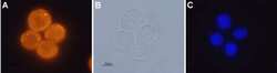
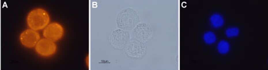
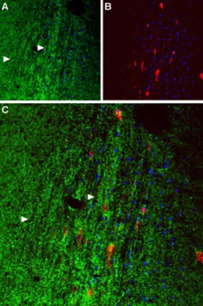
- Immunocytochemistry analysis of Orexin Receptor 1 in paraformaldehyde-fixed and permeabilized human colon cancer cells. A) Samples were probed with Orexin Receptor 1 polyclonal antibody (Product # PA5-77566) at a dilution of 1:200, and incubated with goat-anti-rabbit-AlexaFluor-555 and Hoechst. B) Shows live view of samples. C) Shows nuclei stained image.
Supportive validation
- Submitted by
- Invitrogen Antibodies (provider)
- Main image
- Experimental details
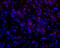
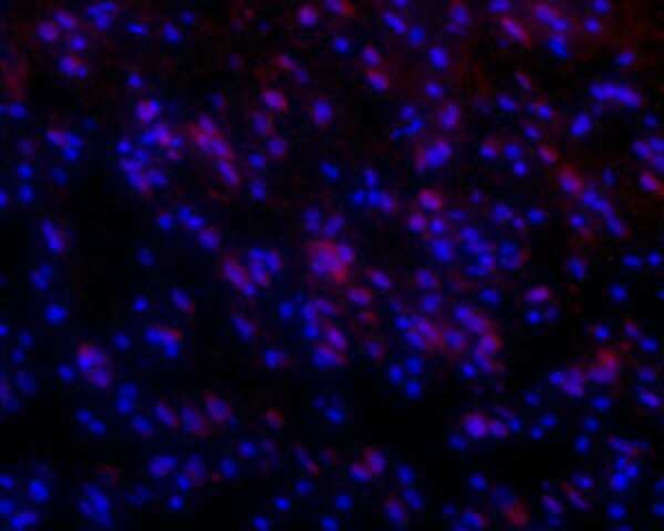
- Immunohistochemistry analysis of Orexin Receptor 1 in frozen rat brainstem. Samples were probed with Orexin Receptor 1 polyclonal antibody (Product # PA5-77566) using a dilution of 1:50, and incubated with goat-anti-rabbit-AlexaFluor-555 and Hoechst.
- Submitted by
- Invitrogen Antibodies (provider)
- Main image
- Experimental details
- Immunohistochemistry analysis of Orexin Receptor 1 in frozen rat brainstem. Samples were probed with Orexin Receptor 1 polyclonal antibody (Product # PA5-77566) using a dilution of 1:50, and incubated with goat-anti-rabbit-AlexaFluor-555 and Hoechst.
- Submitted by
- Invitrogen Antibodies (provider)
- Main image

- Experimental details
- Immunohistochemistry analysis of Orexin Receptor 1 in frozen rat brainstem. Samples were probed with Orexin Receptor 1 polyclonal antibody (Product # PA5-77566) using a dilution of 1:50, and incubated with goat-anti-rabbit-AlexaFluor-555 and Hoechst.
Supportive validation
- Submitted by
- Invitrogen Antibodies (provider)
- Main image
- Experimental details
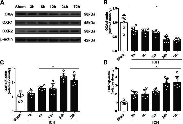
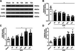
- Fig. 1 Time-course study of OXA, OXR1, and OXR2 using western blot. a Representative protein bands. b Western blot quantitative analyses of OXA, one-way ANOVA, * p < 0.05 vs. sham group. Error bars represent the means +- SD, n = 6 per group. c Western blot quantitative analyses of OXR1, one-way ANOVA, * p < 0.05 vs. sham group. Error bars represent the means +- SD, n = 6 per group. d Western blot quantitative analyses of OXR2, one-way ANOVA, * p < 0.05 vs. sham group. Error bars represent mean +- SD, n = 6 per group