Explore
 Validate
 Learn
Learn Western blot
Antibody data
- Antibody Data
- Antigen structure
- References [0]
- Comments [0]
- Validations
- Western blot [3]
- Immunocytochemistry [1]
- Immunohistochemistry [3]
Submit
Validation data
Reference
Comment
Report error
- Product number
- AOR-001-200UL - Provider product page

- Provider
- Invitrogen Antibodies
- Product name
- Orexin Receptor 1 Polyclonal Antibody
- Antibody type
- Polyclonal
- Antigen
- Other
- Reactivity
- Human, Mouse, Rat
- Host
- Rabbit
- Isotype
- IgG
- Vial size
- 200 µL
- Concentration
- 0.8 mg/mL
- Storage
- -20° C, Avoid Freeze/Thaw Cycles
No comments: Submit comment
Supportive validation
- Submitted by
- Invitrogen Antibodies (provider)
- Main image
- Experimental details
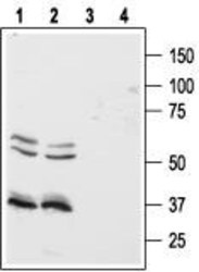
- Western blot analysis of human Colo-205 (lanes 1 and 3) and HT-29 (lanes 2 and 4) colon cancer cell lines: - 1,2. Anti-Orexin Receptor 1 Antibody (#AOR-001), (1:500).3,4. Anti-Orexin Receptor 1 Antibody , preincubated with Orexin Receptor 1 Blocking Peptide (#BLP-OR001).
- Submitted by
- Invitrogen Antibodies (provider)
- Main image
- Experimental details
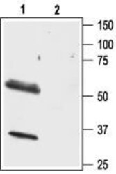
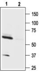
- Western blot analysis of rat brain lysate: - 1. Anti-Orexin Receptor 1 Antibody (#AOR-001), (1:500). 2. Anti-Orexin Receptor 1 Antibody , preincubated with Orexin Receptor 1 Blocking Peptide (#BLP-OR001).

- Submitted by
- Invitrogen Antibodies (provider)
- Main image
- Experimental details
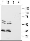
- Western blot analysis of mouse brain lysate: - 1. Anti-Orexin Receptor 1 Antibody (#AOR-001), (1:200). 2. Anti-Orexin Receptor 1 Antibody , preincubated with Orexin Receptor 1 Blocking Peptide (#BLP-OR001).

Supportive validation
- Submitted by
- Invitrogen Antibodies (provider)
- Main image
- Experimental details
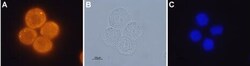
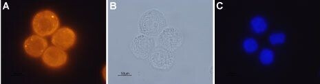
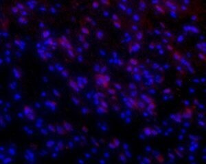
- Expression of OX1R in human colon cancer cell lines - Immunocytochemical staining of paraformaldehyde fixed and permeabilized human Colo-205 colon cancer cells A. Cells were stained with Anti-Orexin Receptor 1 Antibody (#AOR-001), (1:200), followed by goat- Anti-rabbit-AlexaFluor-555 secondary Antibody . B. Live view of the same field as in (A). C. Nuclei were visualized with the cell permeable dye Hoechst 33342 (blue staining).
Supportive validation
- Submitted by
- Invitrogen Antibodies (provider)
- Main image
- Experimental details
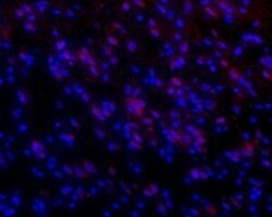
- Expression of OX1R in rat brain - Longitudinal frozen section of rat brainstem showing staining (red) in neuronal cell bodies. Slides were incubated overnight at 4°C with Anti-Orexin Receptor 1 Antibody (#AOR-001), (1:50) followed by goat- Anti-rabbit-AlexaFluor-555 secondary Antibody (1:500). Hoechst 33342is used as a counterstain (blue).
- Submitted by
- Invitrogen Antibodies (provider)
- Main image

- Experimental details
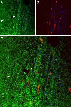
- Expression of OX1R in mouse septum - Immunohistochemical staining paraffin-fixed frozen sections using Anti-Orexin Receptor 1 Antibody (#AOR-001), (1:50). A. OX1R (green) appears in axonal processes (right-pointing triangles). B. Parvalbumin (red) appears in septal neurons. Cell nuclei (blue) are visualized with Hoechst 33342. C. Merge of OX1R and parvalbumin suggests that orexinergic innervation covers the entire septal nucleus rather than restricted to individual neurons.
- Submitted by
- Invitrogen Antibodies (provider)
- Main image
- Experimental details
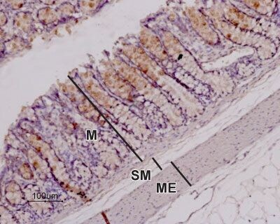
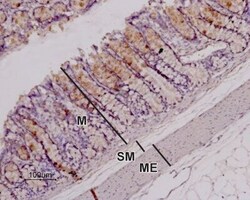
- Expression of OX1R in rat colon - Immunohistochemical staining of paraffin-embedded longitudinal section of rat colon showing mucosa (M), submucosa (SM), andmuscularis externa (ME) using Anti-Orexin Receptor 1 Antibody (#AOR-001), (1:100). Note that the stain (red-brown color) is highly specific for absorptive cells in the superior third of the intestinal glands. Immunolabeling was detected using DAB as the chromogen and hematoxilin as the counterstain.