Explore
 Validate
 Learn
Learn Western blot
 Immunocytochemistry
Antibody data
- Antibody Data
- Antigen structure
- References [8]
- Comments [0]
- Validations
- Immunocytochemistry [2]
Submit
Validation data
Reference
Comment
Report error
- Product number
- HPA039973 - Provider product page

- Provider
- Atlas Antibodies
- Proper citation
- Atlas Antibodies Cat#HPA039973, RRID:AB_10795242
- Product name
- Anti-CPSF6
- Antibody type
- Polyclonal
- Description
- Polyclonal Antibody against Human CPSF6, Gene description: cleavage and polyadenylation specific factor 6, 68kDa, Alternative Gene Names: CFIM, CFIM68, HPBRII-4, HPBRII-7, Validated applications: WB, IHC, ICC, Uniprot ID: Q16630, Storage: Store at +4°C for short term storage. Long time storage is recommended at -20°C.
- Reactivity
- Human, Mouse, Rat
- Host
- Rabbit
- Conjugate
- Unconjugated
- Isotype
- IgG
- Vial size
- 100 µl
- Concentration
- 0.2 mg/ml
- Storage
- Store at +4°C for short term storage. Long time storage is recommended at -20°C.
- Handling
- The antibody solution should be gently mixed before use.
Submitted references HIV-1 Capsid Rapidly Induces Long-Lived CPSF6 Puncta in Non-Dividing Cells, but Similar Puncta Already Exist in Uninfected T-Cells
Direct Capsid Labeling of Infectious HIV-1 by Genetic Code Expansion Allows Detection of Largely Complete Nuclear Capsids and Suggests Nuclear Entry of HIV-1 Complexes via Common Routes.
CRISPR/Cas9-Induced Mutagenesis Corroborates the Role of Transportin-SR2 in HIV-1 Nuclear Import
Cone-shaped HIV-1 capsids are transported through intact nuclear pores
HIV-1 requires capsid remodelling at the nuclear pore for nuclear entry and integration
HIV-1 uncoating by release of viral cDNA from capsid-like structures in the nucleus of infected cells.
HIV-1 nuclear import in macrophages is regulated by CPSF6-capsid interactions at the nuclear pore complex.
Quantitative microscopy of functional HIV post-entry complexes reveals association of replication with the viral capsid
Guedán A, Burley M, Caroe E, Bishop K
Viruses 2024;16(5):670
Viruses 2024;16(5):670
Direct Capsid Labeling of Infectious HIV-1 by Genetic Code Expansion Allows Detection of Largely Complete Nuclear Capsids and Suggests Nuclear Entry of HIV-1 Complexes via Common Routes.
Schifferdecker S, Zila V, Müller TG, Sakin V, Anders-Össwein M, Laketa V, Kräusslich HG, Müller B
mBio 2022 Oct 26;13(5):e0195922
mBio 2022 Oct 26;13(5):e0195922
CRISPR/Cas9-Induced Mutagenesis Corroborates the Role of Transportin-SR2 in HIV-1 Nuclear Import
Janssens J, Blokken J, Lampi Y, De Wit F, Zurnic Bonisch I, Nombela I, Van de Velde P, Van Remoortel B, Gijsbers R, Christ F, Debyser Z, Martinez M
Microbiology Spectrum 2021;9(2)
Microbiology Spectrum 2021;9(2)
Cone-shaped HIV-1 capsids are transported through intact nuclear pores
Zila V, Margiotta E, Turoňová B, Müller T, Zimmerli C, Mattei S, Allegretti M, Börner K, Rada J, Müller B, Lusic M, Kräusslich H, Beck M
Cell 2021;184(4):1032-1046.e18
Cell 2021;184(4):1032-1046.e18
HIV-1 requires capsid remodelling at the nuclear pore for nuclear entry and integration
Swanstrom R, Guedán A, Donaldson C, Caroe E, Cosnefroy O, Taylor I, Bishop K
PLOS Pathogens 2021;17(9):e1009484
PLOS Pathogens 2021;17(9):e1009484
HIV-1 uncoating by release of viral cDNA from capsid-like structures in the nucleus of infected cells.
Müller TG, Zila V, Peters K, Schifferdecker S, Stanic M, Lucic B, Laketa V, Lusic M, Müller B, Kräusslich HG
eLife 2021 Apr 27;10
eLife 2021 Apr 27;10
HIV-1 nuclear import in macrophages is regulated by CPSF6-capsid interactions at the nuclear pore complex.
Bejarano DA, Peng K, Laketa V, Börner K, Jost KL, Lucic B, Glass B, Lusic M, Müller B, Kräusslich HG
eLife 2019 Jan 23;8
eLife 2019 Jan 23;8
Quantitative microscopy of functional HIV post-entry complexes reveals association of replication with the viral capsid
Peng K, Muranyi W, Glass B, Laketa V, Yant S, Tsai L, Cihlar T, Müller B, Kräusslich H
eLife 2014;3
eLife 2014;3
No comments: Submit comment
Enhanced validation
Supportive validation
- Submitted by
- 55af80e3e0991
- Enhanced method
- Genetic validation
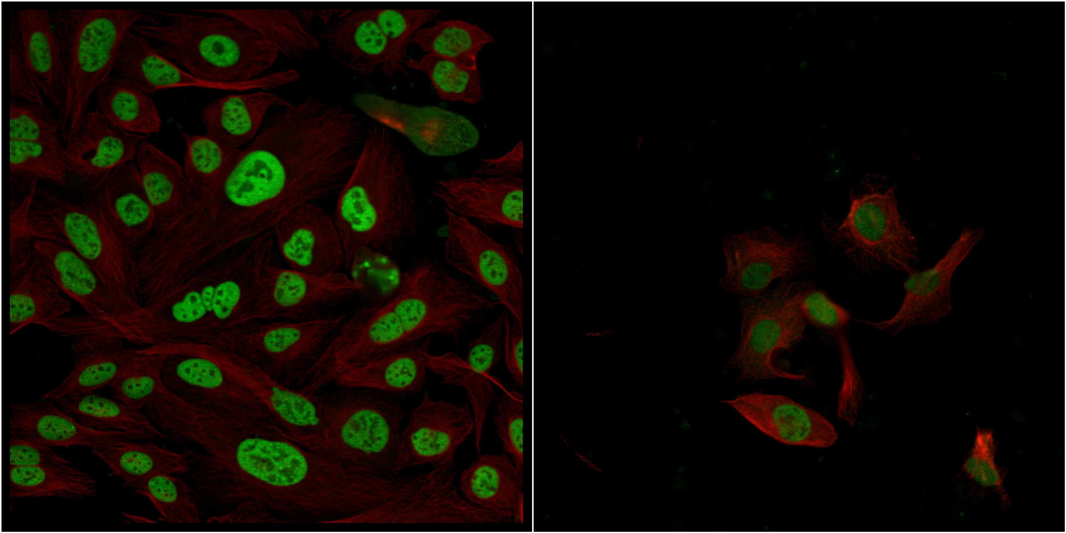
- Main image
- Experimental details
- Confocal images of immunofluorescently stained human U-2 OS cells.The protein CPSF6 is shown in green and the microtubules in red. The image to the left show cells transfected with control siRNA and the image to the right show cells where CPSF6 has been downregulated with specific siRNA.
- Sample type
- U-2 OS cells
- Primary Ab dilution
- 1:74
- Secondary Ab
- Secondary Ab
- Secondary Ab dilution
- 1:800
- Knockdown/Genetic Approaches Application
- Immunocytochemistry
Supportive validation
- Submitted by
- Atlas Antibodies (provider)
- Main image

- Experimental details
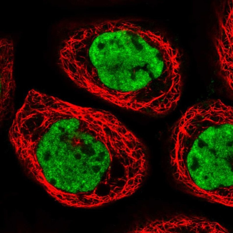
- Immunofluorescent staining of human cell line A-431 shows localization to nucleoplasm & nuclear speckles.
- Sample type
- Human