Explore
 Validate
 Learn
Learn Western blot
Antibody data
- Antibody Data
- Antigen structure
- References [1]
- Comments [0]
- Validations
- Western blot [2]
- Immunocytochemistry [2]
- Immunohistochemistry [4]
Submit
Validation data
Reference
Comment
Report error
- Product number
- PA5-18666 - Provider product page

- Provider
- Invitrogen Antibodies
- Product name
- CGI58 Polyclonal Antibody
- Antibody type
- Polyclonal
- Antigen
- Synthetic peptide
- Description
- This antibody is predicted to react with canine and rat based on sequence homology. This antibody is tested in Peptide ELISA: antibody detection limit dilution 16,000.
- Reactivity
- Human, Mouse
- Host
- Goat
- Isotype
- IgG
- Vial size
- 100 μg
- Concentration
- 0.5 mg/mL
- Storage
- -20°C, Avoid Freeze/Thaw Cycles
Submitted references DGAT enzymes are required for triacylglycerol synthesis and lipid droplets in adipocytes.
Harris CA, Haas JT, Streeper RS, Stone SJ, Kumari M, Yang K, Han X, Brownell N, Gross RW, Zechner R, Farese RV Jr
Journal of lipid research 2011 Apr;52(4):657-67
Journal of lipid research 2011 Apr;52(4):657-67
No comments: Submit comment
Supportive validation
- Submitted by
- Invitrogen Antibodies (provider)
- Main image
- Experimental details
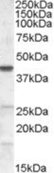
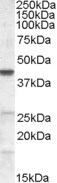
- Western blot analysis of CGI58 using CGI58 Polyclonal Antibody (Product # PA5-18666) (0.2 µg/mL) in staining of NIH3T3 lysate (35 µg protein in RIPA buffer). Detected by chemiluminescence.
- Submitted by
- Invitrogen Antibodies (provider)
- Main image
- Experimental details
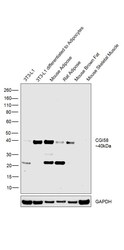
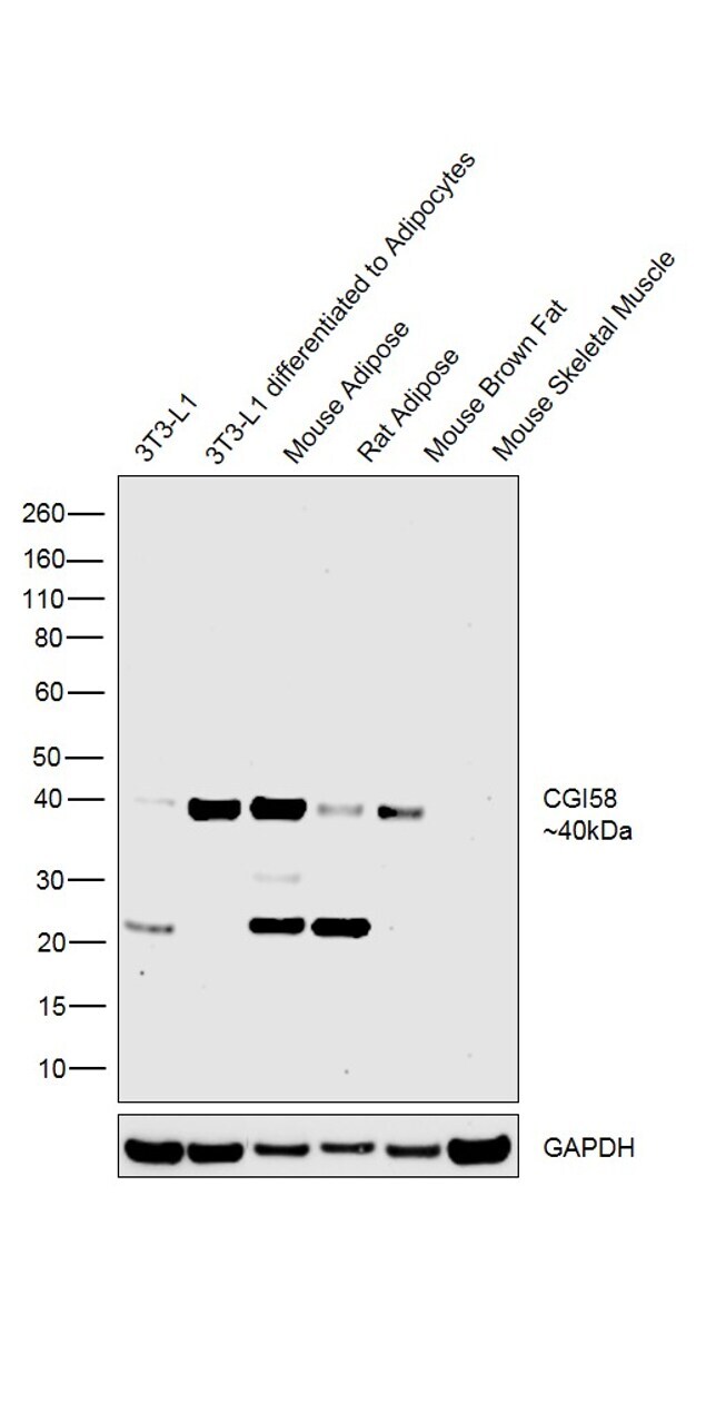
- Western blot was performed using Anti-CGI58 Goat Polyclonal Antibody (Product # PA5-18666) and a 40kDa band corresponding to CGI58 was observed across cell lines and tissues tested except in Mouse Skeletal Muscle which is reported negative for CGI58 expression. An uncharacterized band was also observed at ~22kDa in certain cell and tissue extracts. Whole cell extracts (30 µg lysate) of 3T3-L1 (Lane 1), 3T3-L1 differentiated to adipocytes (Lane 2), Mouse Adipose (Lane 3), Rat Adipose (Lane 4), Mouse Brown Fat (Lane 5) and Mouse Skeletal Muscle (Lane 6) were electrophoresed using Novex® NuPAGE® 4-12 % Bis-Tris gel (Product # NP0322BOX). Resolved proteins were then transferred onto a nitrocellulose membrane (Product # IB23001) by iBlot® 2 Dry Blotting System (Product # IB21001). The blot was probed with the primary antibody (0.5ug/ml) and detected by chemiluminescence Rabbit Anti-Goat IgG Secondary Antibody, HRP conjugate (Product # A27014, 1:4000 dilution) using the iBright FL 1000 (Product # A32752). Chemiluminescent detection was performed using Novex® ECL Chemiluminescent Substrate Reagent Kit (Product # WP20005).
Supportive validation
- Submitted by
- Invitrogen Antibodies (provider)
- Main image

- Experimental details
- Immunofluorescence analysis of CGI58 in U2OS cells using a CGI58 monoclonal antibody (Product # PA5-18666) at 10 µg/mL for1hr. The cells were paraformaldehyde fixed and permeabilized with 0.15% Triton. Primary incubation was followed by Alexa Fluor 488 secondary antibody (2 µg/mL) showing nuclear and vesicle/ cytoplasmic staining. The nuclear stain is DAPI (blue). Negative control: Unimmunized goat IgG (10 µg/mL)followed by Alexa Fluor 488 secondary antibody (2 µg/mL).

- Submitted by
- Invitrogen Antibodies (provider)
- Main image

- Experimental details
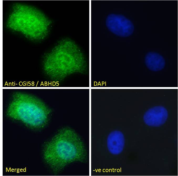
- Immunocytochemistry analysis of CGI58 using CGI58 Polyclonal Antibody (Product # PA5-18666) in paraformaldehyde fixed U2OS cells, permeabilized with 0.15% Triton. Primary incubation 1hr (10 µg/mL) followed by Alexa Fluor 488 secondary antibody (2 µg/mL), showing nuclear and vesicle/ cytoplasmic staining. The nuclear stain is DAPI (blue). Negative control: Unimmunized goat IgG (10 µg/mL) followed by Alexa Fluor 488 secondary antibody (2 µg/mL).
Supportive validation
- Submitted by
- Invitrogen Antibodies (provider)
- Main image
- Experimental details
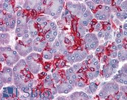
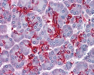
- Immunohistochemistry analysis of CGI58 in human pancreas. Samples were incubated with CGI58 polyclonal antibody (Product # PA5-18666) using a dilution of 3.75 µg/mL. Formalin-fixed, paraffin-embedded tissue after heat-induced antigen retrieval.
- Submitted by
- Invitrogen Antibodies (provider)
- Main image
- Experimental details
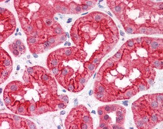
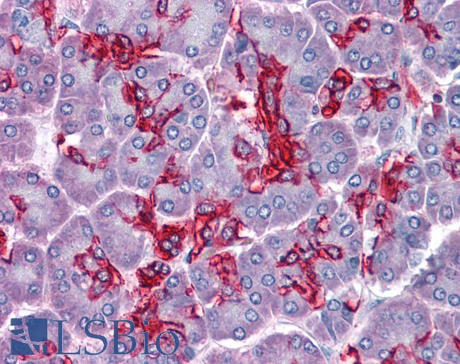
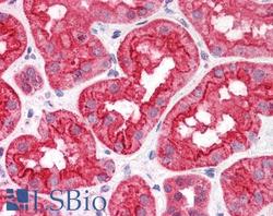
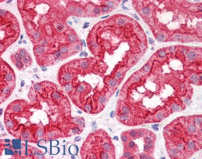
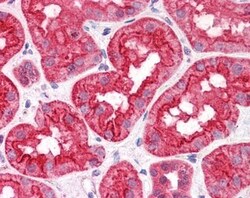
- Immunohistochemistry analysis of CGI58 in human kidney. Samples were incubated with CGI58 polyclonal antibody (Product # PA5-18666) using a dilution of 3.75 µg/mL. Formalin-fixed, paraffin-embedded tissue after heat-induced antigen retrieval.
- Submitted by
- Invitrogen Antibodies (provider)
- Main image

- Experimental details
- Immunohistochemistry analysis of CGI58 in human pancreas. Samples were incubated with CGI58 polyclonal antibody (Product # PA5-18666) using a dilution of 3.75 µg/mL. Formalin-fixed, paraffin-embedded tissue after heat-induced antigen retrieval.
- Submitted by
- Invitrogen Antibodies (provider)
- Main image
- Experimental details
- Immunohistochemistry analysis of CGI58 in human kidney. Samples were incubated with CGI58 polyclonal antibody (Product # PA5-18666) using a dilution of 3.75 µg/mL. Formalin-fixed, paraffin-embedded tissue after heat-induced antigen retrieval.