Explore
 Validate
 Learn
Learn Western blot
Antibody data
- Antibody Data
- Antigen structure
- References [0]
- Comments [0]
- Validations
- Western blot [1]
- Immunocytochemistry [1]
- Immunohistochemistry [1]
- Flow cytometry [1]
Submit
Validation data
Reference
Comment
Report error
- Product number
- AMM85978 - Provider product page

- Provider
- EnkiLife Biotech Co., Ltd.
- Product name
- ATG4A Mouse Monoclonal Antibody
- Antibody type
- Monoclonal
- Description
- Affinity Purification
- Reactivity
- Human
- Host
- Mouse
- Conjugate
- Unconjugated
- Antibody clone number
- Monoclonal
- Vial size
- 100 µl
- Concentration
- 1 mg/ml
- Storage
- Store at 4°C short term. Aliquot and store at -20°C long term. Avoid freeze/thaw cycles.
- Handling
- The antibody solution should be gently mixed before use.
No comments: Submit comment
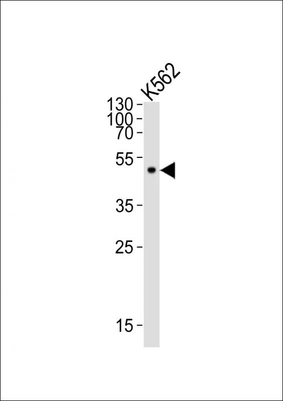
Supportive validation
- Submitted by
- EnkiLife Biotech Co., Ltd. (provider)
- Main image
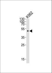
- Experimental details
- Western blot analysis of lysate from K562 cell line, using ATG4A Antibody. ATG4A Mouse Monoclonal Antibody was diluted at 1:500. A goat anti-mouse IgG H&L(HRP) at 1:10000 dilution was used as the secondary antibody. Lysate at 20μg.
Supportive validation
- Submitted by
- EnkiLife Biotech Co., Ltd. (provider)
- Main image
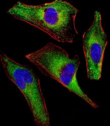
- Experimental details
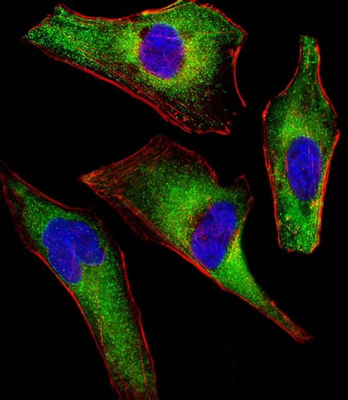
- Immunofluorescent analysis of 4% paraformaldehyde-fixed, 0.1% Triton X-100 permeabilized HeLa (human cervical epithelial adenocarcinoma cell line) cells labeling ATG4A with AMM85978 at 1/25 dilution, followed by Dylight® 488-conjugated goat anti-mouse IgG secondary antibody at 1/200 dilution (green). Immunofluorescence image showing cytoplasm staining on HeLa cell line. Cytoplasmic actin is detected with Dylight® 554 Phalloidin at 1/100 dilution (red).The nuclear counter stain is DAPI (blue).
Supportive validation
- Submitted by
- EnkiLife Biotech Co., Ltd. (provider)
- Main image
- Experimental details
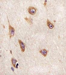
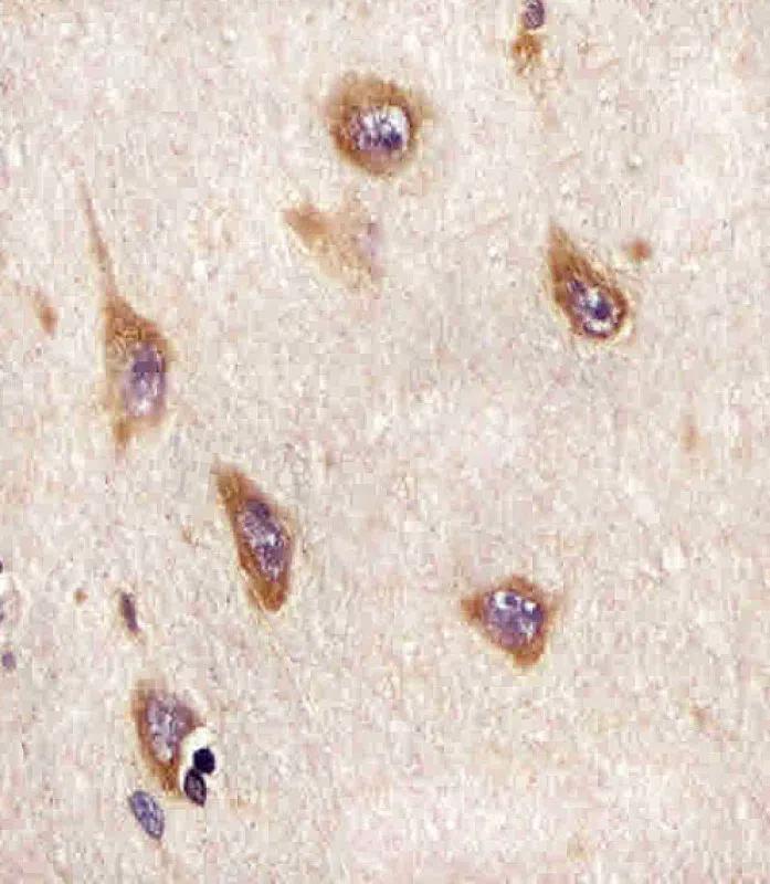
- AMM85978 staining ATG4A in human brain sections by Immunohistochemistry (IHC-P - paraformaldehyde-fixed, paraffin-embedded sections). Tissue was fixed with formaldehyde and blocked with 3% BSA for 0. 5 hour at room temperature; antigen retrieval was by heat mediation with a citrate buffer (pH6). Samples were incubated with primary antibody (1/25) for 1 hours at 37°C. A undiluted biotinylated goat polyvalent antibody was used as the secondary antibody.
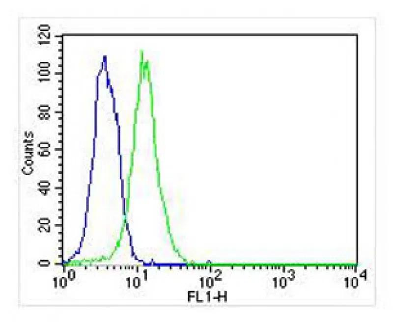
Supportive validation
- Submitted by
- EnkiLife Biotech Co., Ltd. (provider)
- Main image
- Experimental details
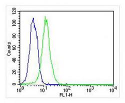
- Overlay histogram showing Hela cells stained with AMM85978 (green line). The cells were fixed with 2% paraformaldehyde (10 min) and then permeabilized with 90% methanol for 10 min. The cells were then icubated in 2% bovine serum albumin to block non-specific protein-protein interactions followed by the antibody (AMM85978, 1:25 dilution) for 60 min at 37ºC. The secondary antibody used was Goat-Anti-Mouse IgG, DyLight® 488 Conjugated Highly Cross-Adsorbed(NA168821)) at 1/400 dilution for 40 min at 37ºC. Isotype control antibody (blue line) was mouse IgG2b (1μg/1x10^6 cells) used under the same conditions. Acquisition of >10, 000 events was performed.