Explore
 Validate
 Learn
Learn Western blot
 Immunocytochemistry
Antibody data
- Antibody Data
- Antigen structure
- References [4]
- Comments [0]
- Validations
- Immunocytochemistry [2]
- Immunohistochemistry [3]
- Other assay [1]
Submit
Validation data
Reference
Comment
Report error
- Product number
- PA3-065 - Provider product page

- Provider
- Invitrogen Antibodies
- Product name
- Endothelin A Receptor Polyclonal Antibody
- Antibody type
- Polyclonal
- Antigen
- Other
- Description
- PA3-065 detects Endothelin Receptor A from human, mouse, and rat samples. PA3-065 has been successfully used in Western blot, immunocytochemistry, and immunohistochemistry procedures. By western blot, this antibody detects a 46kDa protein corresponding to mouse Endothelin Receptor A.
- Reactivity
- Human, Mouse, Rat
- Host
- Rabbit
- Isotype
- IgG
- Vial size
- 100 μL
- Concentration
- Conc. Not Determined
- Storage
- -20°C, Avoid Freeze/Thaw Cycles
Submitted references Endothelin-1 induces changes in the expression levels of steroidogenic enzymes and increases androgen receptor and testosterone production in the PC3 prostate cancer cell line.
Peroxisome proliferator-activated receptor gamma blunts endothelin-1-mediated contraction of the uterine artery in a murine model of high-altitude pregnancy.
Reassessment of endothelin receptor A expression in normal and neoplastic human tissues using the novel rabbit monoclonal antibody UMB-8.
Transforming growth factor-β regulates endothelin-1 signaling in the newborn mouse lung during hypoxia exposure.
Torres MJ, López-Moncada F, Herrera D, Indo S, Lefian A, Llanos P, Tapia J, Castellón EA, Contreras HR
Oncology reports 2021 Aug;46(2)
Oncology reports 2021 Aug;46(2)
Peroxisome proliferator-activated receptor gamma blunts endothelin-1-mediated contraction of the uterine artery in a murine model of high-altitude pregnancy.
Lane SL, Doyle AS, Bales ES, Houck JA, Lorca RA, Moore LG, Julian CG
FASEB journal : official publication of the Federation of American Societies for Experimental Biology 2020 Mar;34(3):4283-4292
FASEB journal : official publication of the Federation of American Societies for Experimental Biology 2020 Mar;34(3):4283-4292
Reassessment of endothelin receptor A expression in normal and neoplastic human tissues using the novel rabbit monoclonal antibody UMB-8.
Lupp A, Mann A, Heeb A, Kaemmerer D, Sänger J, Evert M, Evert K, Mawrin C, Schulz S
Peptides 2015 Apr;66:19-25
Peptides 2015 Apr;66:19-25
Transforming growth factor-β regulates endothelin-1 signaling in the newborn mouse lung during hypoxia exposure.
Olave N, Nicola T, Zhang W, Bulger A, James M, Oparil S, Chen YF, Ambalavanan N
American journal of physiology. Lung cellular and molecular physiology 2012 May 1;302(9):L857-65
American journal of physiology. Lung cellular and molecular physiology 2012 May 1;302(9):L857-65
No comments: Submit comment
Supportive validation
- Submitted by
- Invitrogen Antibodies (provider)
- Main image

- Experimental details
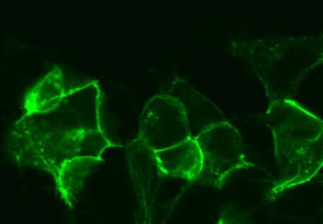
- Immunofluorescent analysis of Endothelin A Receptor in ETA-transfected cells using a polyclonal antibody (Product # PA3-065).

- Submitted by
- Invitrogen Antibodies (provider)
- Main image

- Experimental details
- Immunofluorescent analysis of Endothelin A Receptor in ETA-transfected cells using a polyclonal antibody (Product # PA3-065).
Supportive validation
- Submitted by
- Invitrogen Antibodies (provider)
- Main image

- Experimental details
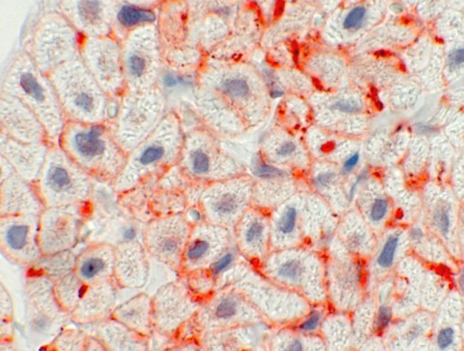
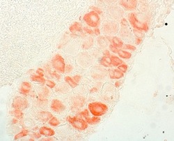
- Immunohistochemistry of ETA receptors in the atrium of mouse heart.
- Submitted by
- Invitrogen Antibodies (provider)
- Main image
- Experimental details
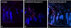
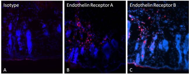
- Immunohistochemistry was performed on acetone-fixed frozen mouse skin tissue sections. Tissues were fixed for 10 minutes, blocked in 3% fish gelatin for 10-20 minutes, and probed with an Isotype Control Antibody (panel A), and Endothelin Receptor A polyclonal antibody (Product # PA3-065, panel B), or an Endothelin Receptor B polyclonal antibody (Product # PA3-066, panel C) all at dilutions of 1:800 overnight at 4C. Detection was performed using a PE-conjugated anti-rabbit IgG secondary antibody at a dilution of 1:800 (red). Nuclei were stained with DAPI and tissues were visualized by fluorescence microscopy. Data courtesy of the Innovators Program.
- Submitted by
- Invitrogen Antibodies (provider)
- Main image
- Experimental details
- Immunohistochemistry of ETA in mouse dorsal root ganglia

Supportive validation
- Submitted by
- Invitrogen Antibodies (provider)
- Main image
- Experimental details
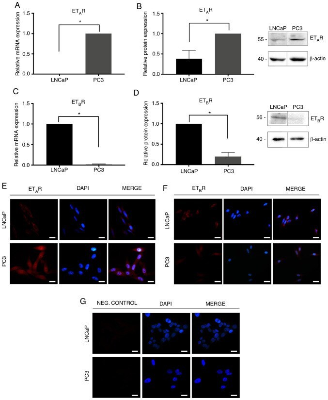
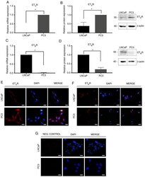
- Figure 2. ET A R and ET B R expression in prostate cancer cells. (A) ET A R mRNA expression normalized to Pumilio compared with PC3 cells. (B) ET A R protein expression in PC3 and LNCaP cells. Quantification was normalized to beta-actin and PC3 cells. (C) ET B R mRNA expression normalized to Pumilio compared with LNCaP cells. (D) ET B R protein expression in PC3 and LNCaP cells normalized to Pumilio compared with LNCaP cells. Immunofluorescence analysis of (E) ET A R and (F) ET B R in PC3 and LNCaP cells. (G) Negative controls. Data are presented as the mean +- SD (n=3 independent experiments) of arbitrary units. *P