 Explore
 Validate
 Learn
Learn10494-1-AP
antibody from Invitrogen Antibodies
Targeting: GAPDHS
GAPD2, GAPDH-2, GAPDS
 Western blot Immunocytochemistry
 Immunoprecipitation Immunohistochemistry Flow cytometry Other assay
Antibody data
- Antibody Data
- Antigen structure
- References [0]
- Comments [0]
- Validations
- Western blot [12]
- Immunocytochemistry [3]
- Immunohistochemistry [6]
- Flow cytometry [1]
- Other assay [1]
Submit
Validation data
Reference
Comment
Report error
- Product number
- 10494-1-AP - Provider product page

- Provider
- Invitrogen Antibodies
- Product name
- GAPDH Polyclonal Antibody
- Antibody type
- Polyclonal
- Antigen
- Other
- Reactivity
- Human, Mouse, Rat, Porcine
- Host
- Rabbit
- Isotype
- IgG
- Vial size
- 150 µL
- Concentration
- 0.33 mg/mL
- Storage
- -20°C
No comments: Submit comment
Supportive validation
- Submitted by
- Invitrogen Antibodies (provider)
- Main image

- Experimental details
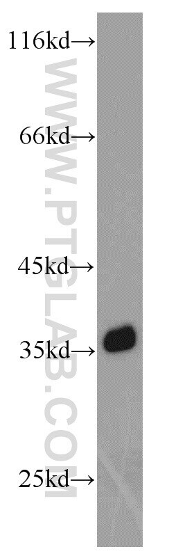
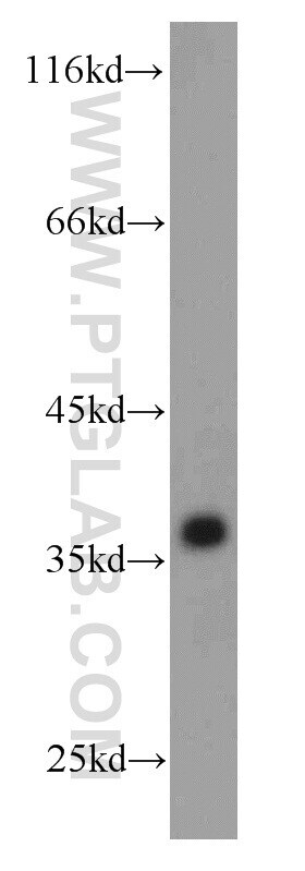
- HEK-293 cells were subjected to SDS PAGE followed by western blot with 10494-1-AP (GAPDH Antibody) at dilution of 1:5000 incubated at room temperature for 1.5 hours.
- Submitted by
- Invitrogen Antibodies (provider)
- Main image
- Experimental details
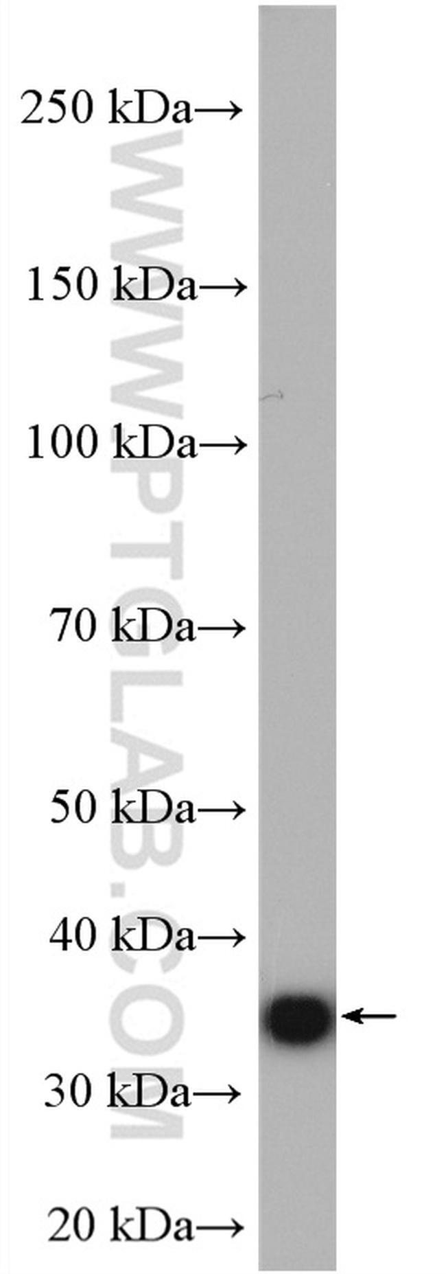
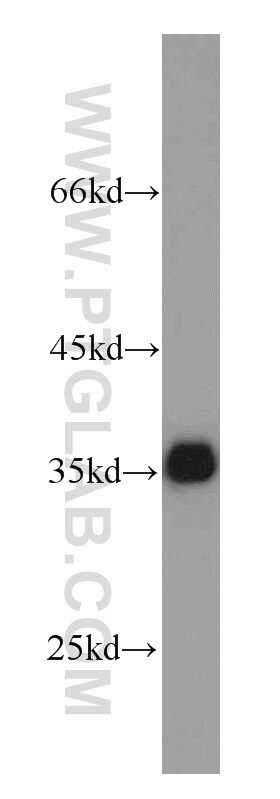
- HeLa cells were subjected to SDS PAGE followed by western blot with 10494-1-AP (GAPDH antibody) at dilution of 1:20000 incubated at room temperature for 1.5 hours.
- Submitted by
- Invitrogen Antibodies (provider)
- Main image
- Experimental details
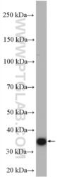
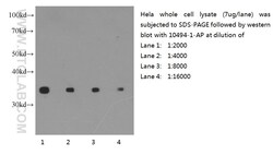
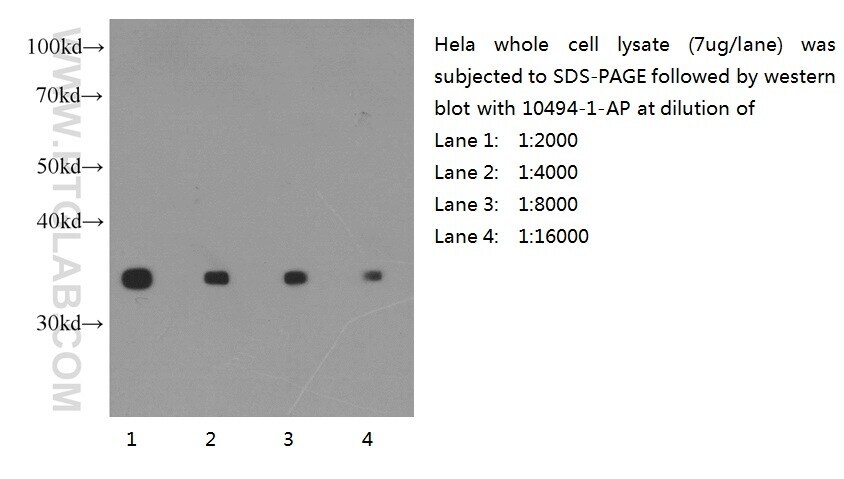
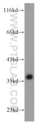
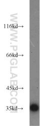
- Western blot of Hela cell with anti-GAPDH (10494-1-AP) at various dilutions.
- Submitted by
- Invitrogen Antibodies (provider)
- Main image
- Experimental details
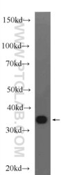
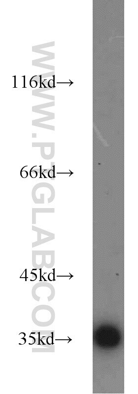
- Arabidopsis whole plant tissue were subjected to SDS PAGE followed by western blot with 10494-1-AP (GAPDH antibody) at dilution of 1:3000 incubated at room temperature for 1.5 hours.

- Submitted by
- Invitrogen Antibodies (provider)
- Main image
- Experimental details
- Corn whole plant tissue were subjected to SDS PAGE followed by western blot with 10494-1-AP (GAPDH antibody) at dilution of 1:3000 incubated at room temperature for 1.5 hours.

- Submitted by
- Invitrogen Antibodies (provider)
- Main image

- Experimental details
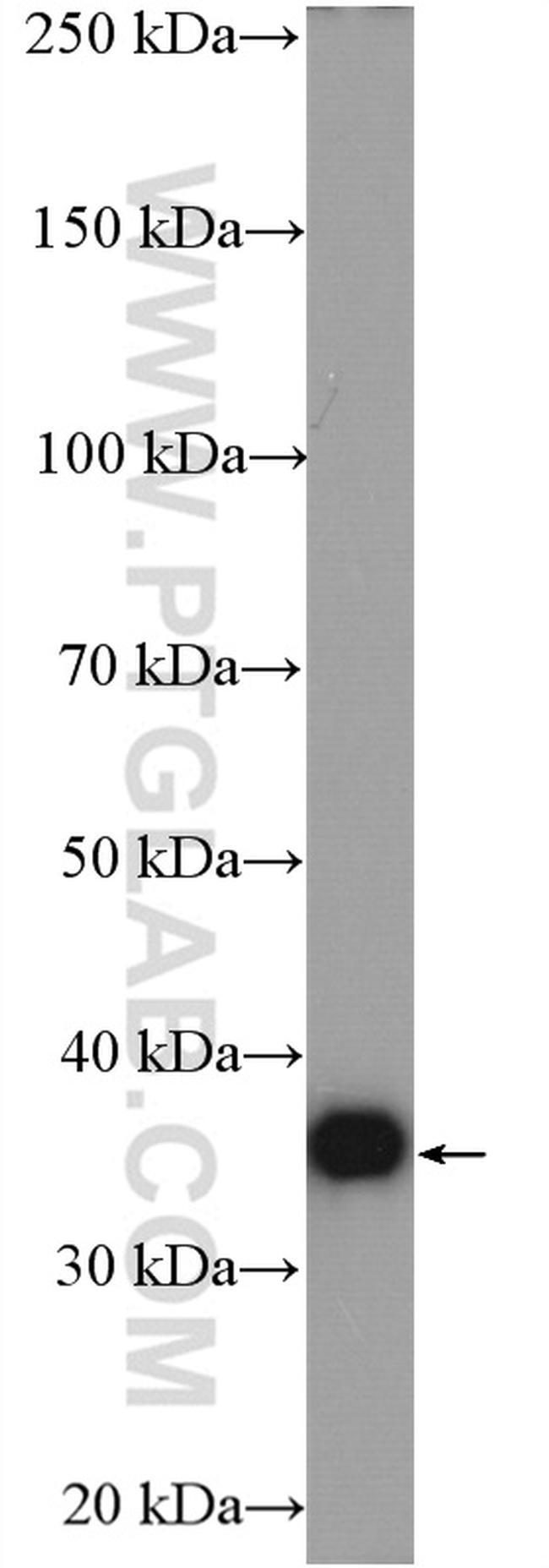
- Mouse brain tissue were subjected to SDS PAGE followed by western blot with 10494-1-AP (GAPDH Antibody) at dilution of 1:20000 incubated at room temperature for 1.5 hours.
- Submitted by
- Invitrogen Antibodies (provider)
- Main image
- Experimental details
- Rat brain tissue were subjected to SDS PAGE followed by western blot with 10494-1-AP (GAPDH Antibody) at dilution of 1:3000 incubated at room temperature for 1.5 hours.

- Submitted by
- Invitrogen Antibodies (provider)
- Main image

- Experimental details
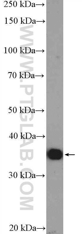
- HEK-293 cells were subjected to SDS PAGE followed by western blot with 10494-1-AP (GAPDH antibody) at dilution of 1:2000 incubated at room temperature for 1.5 hours.
- Submitted by
- Invitrogen Antibodies (provider)
- Main image

- Experimental details
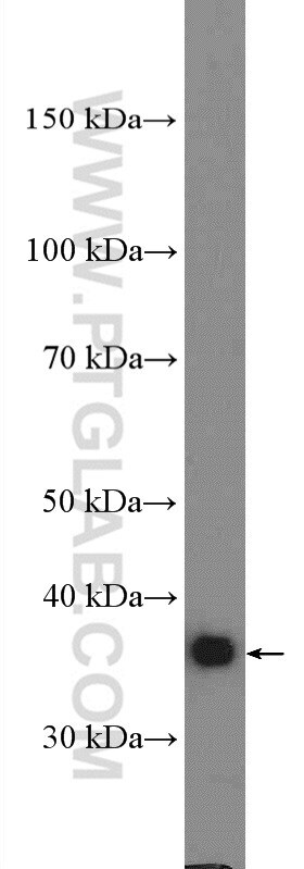
- Raji cells were subjected to SDS PAGE followed by western blot with 10494-1-AP (GAPDH antibody) at dilution of 1:5000 incubated at room temperature for 1.5 hours.
- Submitted by
- Invitrogen Antibodies (provider)
- Main image
- Experimental details
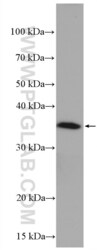
- K-562 cells were subjected to SDS PAGE followed by western blot with 10494-1-AP (GAPDH antibody) at dilution of 1:5000 incubated at room temperature for 1.5 hours.
- Submitted by
- Invitrogen Antibodies (provider)
- Main image

- Experimental details
- HeLa cells were subjected to SDS PAGE followed by western blot with 10494-1-AP (GAPDH antibody) at dilution of 1:5000 incubated at room temperature for 1.5 hours.
- Submitted by
- Invitrogen Antibodies (provider)
- Main image
- Experimental details
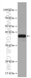
- A549 cells were subjected to SDS PAGE followed by western blot with 10494-1-AP (GAPDH antibody) at dilution of 1:3000 incubated at room temperature for 1.5 hours.
Supportive validation
- Submitted by
- Invitrogen Antibodies (provider)
- Main image
- Experimental details
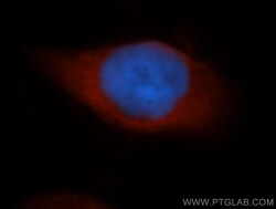
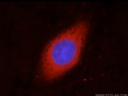
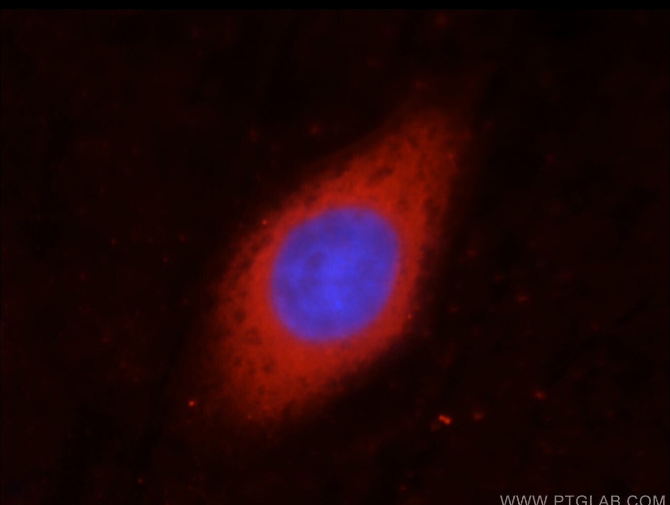
- Immunofluorescent analysis of Hela cells, using GAPDH antibody 10494-1-AP at 1:50 dilution and Rhodamine-labeled goat anti-rabbit IgG (red). Blue pseudocolor = DAPI (fluorescent DNA dye).
- Submitted by
- Invitrogen Antibodies (provider)
- Main image
- Experimental details
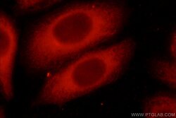
- Immunofluorescent analysis of HepG2 cells, using GAPDH antibody 10494-1-AP at 1:50 dilution and Rhodamine-labeled goat anti-rabbit IgG (red).

- Submitted by
- Invitrogen Antibodies (provider)
- Main image
- Experimental details
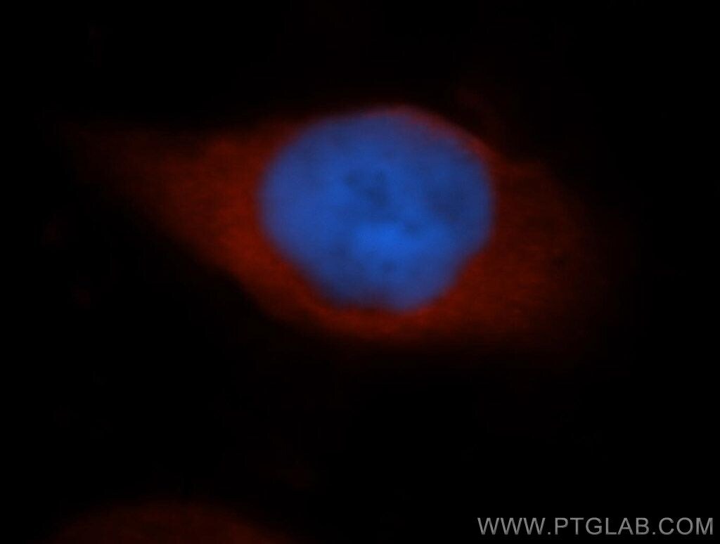
- Immunofluorescent analysis of HepG2 cells, using GAPDH antibody 10494-1-AP at 1:50 dilution and Rhodamine-labeled goat anti-rabbit IGG (red). Blue pseudocolor = DAPI (fluorescent DNA dye).
Supportive validation
- Submitted by
- Invitrogen Antibodies (provider)
- Main image
- Experimental details
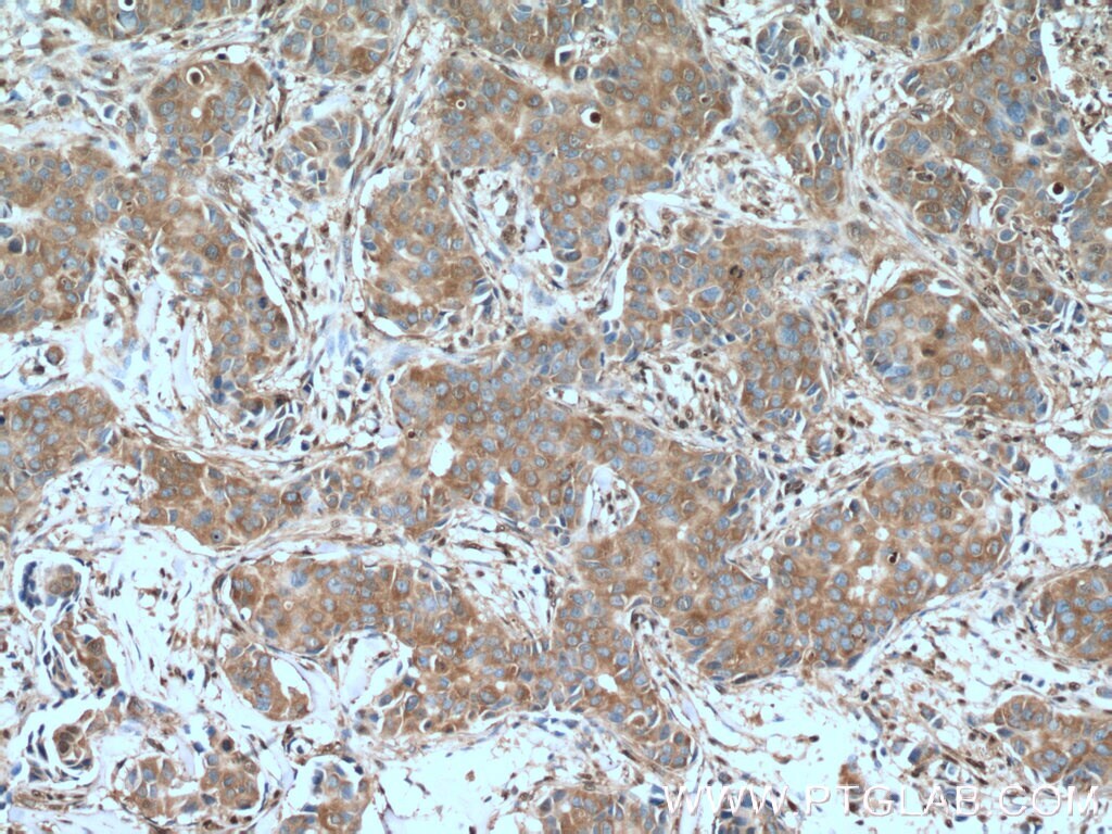
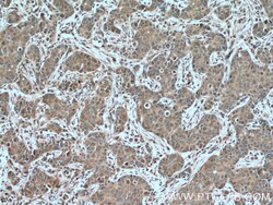
- Immunohistochemistry of paraffin-embedded human breast cancer tissue slide using 10494-1-AP (GAPDH antibody) at dilution of 1:400 (under 10x lens) heat mediated antigen retrieved with Tris-EDTA buffer (pH 9).
- Submitted by
- Invitrogen Antibodies (provider)
- Main image
- Experimental details
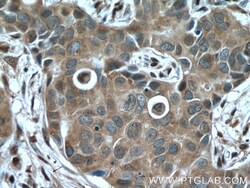
- Immunohistochemistry of paraffin-embedded human breast cancer tissue slide using 10494-1-AP (GAPDH antibody) at dilution of 1:400 (under 40x lens) heat mediated antigen retrieved with Tris-EDTA buffer (pH 9).
- Submitted by
- Invitrogen Antibodies (provider)
- Main image
- Experimental details
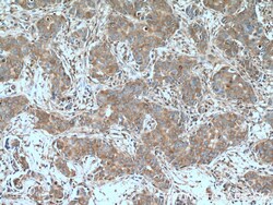
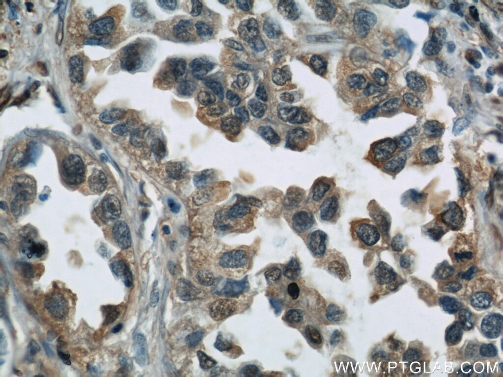
- Immunohistochemistry of paraffin-embedded human lung cancer using 10494-1-AP (GAPDH antibody) at dilution of 1:50 (under 10x lens).
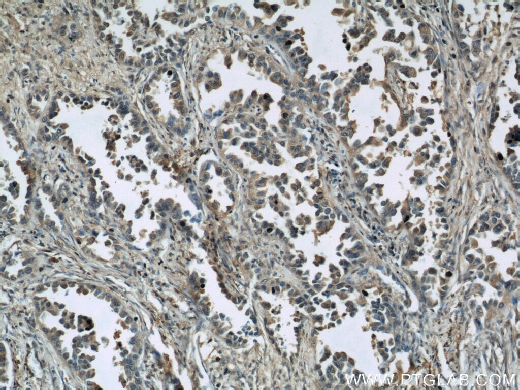
- Submitted by
- Invitrogen Antibodies (provider)
- Main image
- Experimental details
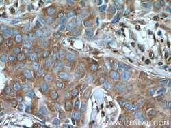
- Immunohistochemistry of paraffin-embedded human lung cancer using 10494-1-AP (GAPDH antibody) at dilution of 1:50 (under 40x lens).
- Submitted by
- Invitrogen Antibodies (provider)
- Main image
- Experimental details
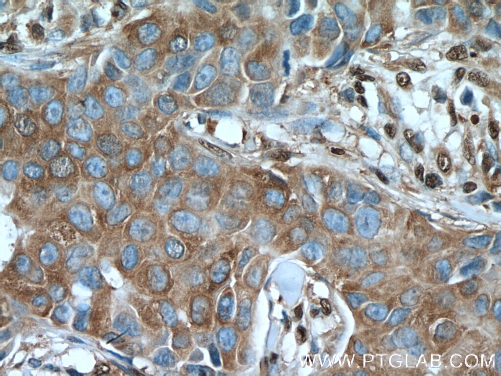
- Immunohistochemistry of paraffin-embedded human breast cancer tissue slide using 10494-1-AP ( GAPDH antibody) at dilution of 1:400 (under 10x lens) heat mediated antigen retrieved with Tris-EDTA buffer (pH 9).

- Submitted by
- Invitrogen Antibodies (provider)
- Main image
- Experimental details
- Immunohistochemistry of paraffin-embedded human breast cancer tissue slide using 10494-1-AP ( GAPDH antibody) at dilution of 1:400 (under 40x lens) heat mediated antigen retrieved with Tris-EDTA buffer (pH 9).

Supportive validation
- Submitted by
- Invitrogen Antibodies (provider)
- Main image
- Experimental details
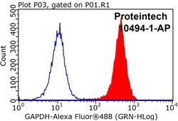
- 1X10^6 HEK-293 cells were stained with .2ug GAPDH antibody (10494-1-AP, red) and control antibody (blue). Fixed with 90% MeOH blocked with 3% BSA (30 min). Alexa Fluor 488 -Goat anti-Rabbit IGG with dilution 1:100.

Supportive validation
- Submitted by
- Invitrogen Antibodies (provider)
- Main image
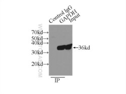
- Experimental details
- IP result of anti-GAPDH (IP:10494-1-AP, 3ug; Detection:10494-1-AP 1:3000) with A549 cells lysate 3500ug.
