Explore
 Validate
 Learn
Learn Western blot
Antibody data
- Antibody Data
- Antigen structure
- References [0]
- Comments [0]
- Validations
- Western blot [1]
- Immunohistochemistry [1]
- Flow cytometry [2]
Submit
Validation data
Reference
Comment
Report error
- Product number
- AF6695 - Provider product page

- Provider
- R&D Systems
- Product name
- Mouse/Rat DARC Antibody
- Antibody type
- Polyclonal
- Description
- Antigen Affinity-purified. Detects mouse and rat DARC in Western blots.
- Reactivity
- Mouse, Rat
- Host
- Sheep
- Conjugate
- Unconjugated
- Antigen sequence
NP_034175- Isotype
- IgG
- Vial size
- 100 ug
- Concentration
- LYOPH
- Storage
- Use a manual defrost freezer and avoid repeated freeze-thaw cycles. 12 months from date of receipt, -20 to -70 °C as supplied. 1 month, 2 to 8 °C under sterile conditions after reconstitution. 6 months, -20 to -70 °C under sterile conditions after reconstitution.
No comments: Submit comment
Supportive validation
- Submitted by
- R&D Systems (provider)
- Main image
- Experimental details
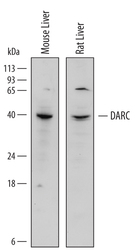
- Detection of Mouse and Rat DARC by Western Blot. Western blot shows lysates of mouse liver tissue and rat liver tissue. PVDF Membrane was probed with 1 µg/mL of Sheep Anti-Mouse/Rat DARC Antigen Affinity-purified Polyclonal Antibody (Catalog # AF6695) followed by HRP-conjugated Anti-Sheep IgG Secondary Antibody (Catalog # HAF016). A specific band was detected for DARC at approximately 40 kDa (as indicated). This experiment was conducted under reducing conditions and using Immunoblot Buffer Group 1.
Supportive validation
- Submitted by
- R&D Systems (provider)
- Main image
- Experimental details
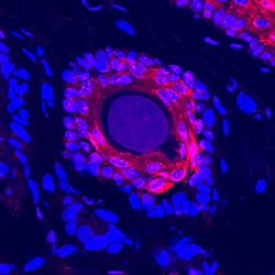
- DARC in Mouse Skin. DARC was detected in perfusion fixed frozen sections of mouse skin using Sheep Anti-Mouse/Rat DARC Antigen Affinity-purified Polyclonal Antibody (Catalog # AF6695) at 0.5 µg/mL overnight at 4 °C. Tissue was stained using the NorthernLights™ 557-conjugated Anti-Sheep IgG Secondary Antibody (red; Catalog # NL010) and counterstained with DAPI (blue). Specific staining was localized to the hair follicle. View our protocol for Fluorescent IHC Staining of Frozen Tissue Sections.

Supportive validation
- Submitted by
- R&D Systems (provider)
- Main image

- Experimental details
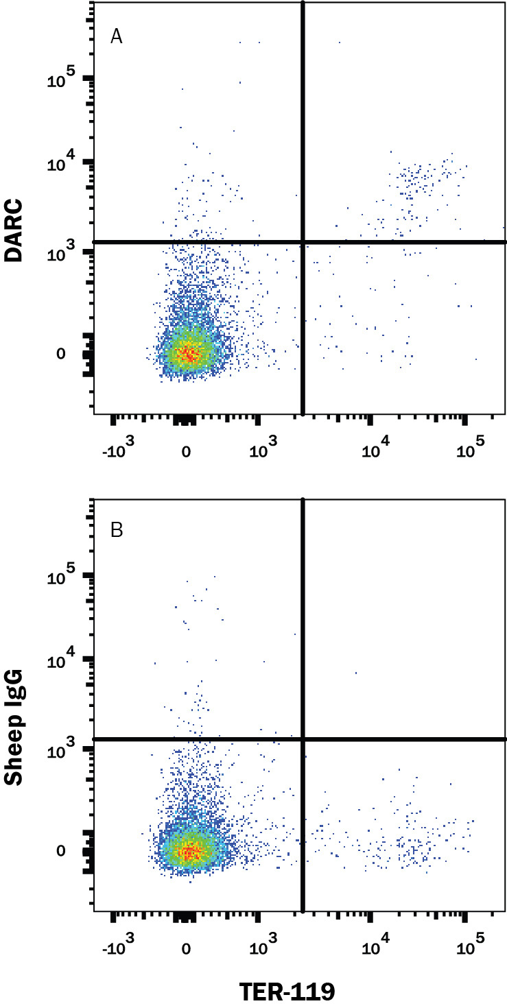
- Detection of DARC in Mouse Splenocytes by Flow Cytometry. Mouse splenocytes were stained with Rat Anti-Mouse TER-119 APC-conjugated Monoclonal Antibody (Catalog # FAB1125A) and either (A) Sheep Anti-Mouse/Rat DARC Antigen Affinity-purified Polyclonal Antibody (Catalog # AF6695) or (B) Normal Sheep IgG Control (Catalog # 5-001-A) followed by Allophycocyanin-conjugated Anti-Sheep IgG Secondary Antibody (Catalog # F0127).
- Submitted by
- R&D Systems (provider)
- Main image

- Experimental details
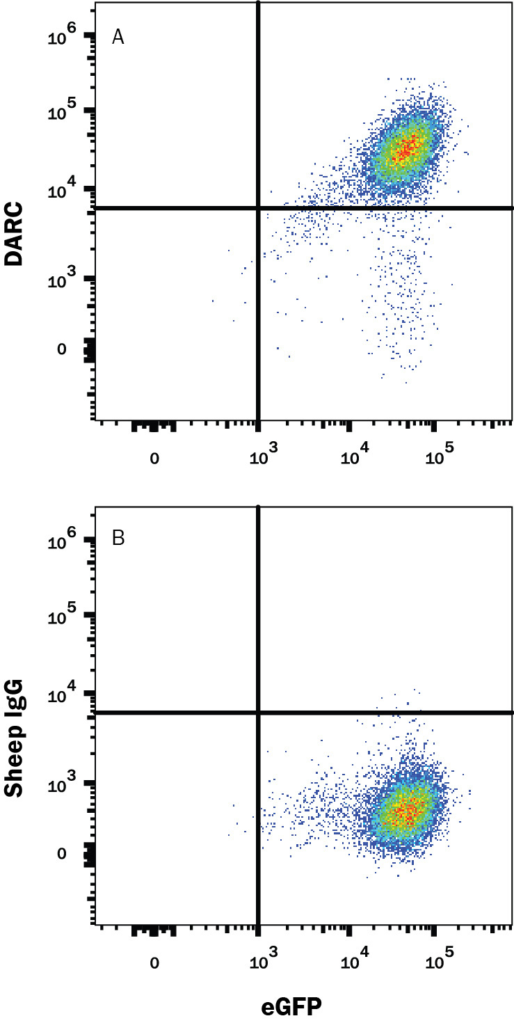
- Detection of DARC in HEK293 Human Cell Line Transfected with Mouse DARC and eGFP by Flow Cytometry. HEK293 human embryonic kidney cell line transfected with mouse DARC and eGFP was stained with either (A) Sheep Anti-Mouse/Rat DARC Antigen Affinity-purified Polyclonal Antibody (Catalog # AF6695) or (B) Normal Sheep IgG Control (Catalog # 5-001-A) followed by Allophycocyanin-conjugated Anti-Sheep IgG Secondary Antibody (Catalog # F0127).