Explore
 Validate
 Learn
Learn Western blot
 Immunocytochemistry
Antibody data
- Antibody Data
- Antigen structure
- References [1]
- Comments [0]
- Validations
- Immunocytochemistry [3]
- Immunohistochemistry [2]
- Other assay [2]
Submit
Validation data
Reference
Comment
Report error
- Product number
- PA5-106850 - Provider product page

- Provider
- Invitrogen Antibodies
- Product name
- NPY5R Polyclonal Antibody
- Antibody type
- Polyclonal
- Antigen
- Synthetic peptide
- Description
- Antibody detects endogenous levels of total NPY5R.
- Reactivity
- Human, Mouse, Rat
- Host
- Rabbit
- Isotype
- IgG
- Vial size
- 100 μL
- Concentration
- 1 mg/mL
- Storage
- -20°C
Submitted references The Novel Methylation Biomarker NPY5R Sensitizes Breast Cancer Cells to Chemotherapy.
Liu J, Wang X, Sun J, Chen Y, Li J, Huang J, Du H, Gan L, Qiu Z, Li H, Ren G, Wei Y
Frontiers in cell and developmental biology 2021;9:798221
Frontiers in cell and developmental biology 2021;9:798221
No comments: Submit comment
Supportive validation
- Submitted by
- Invitrogen Antibodies (provider)
- Main image
- Experimental details
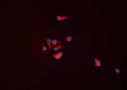
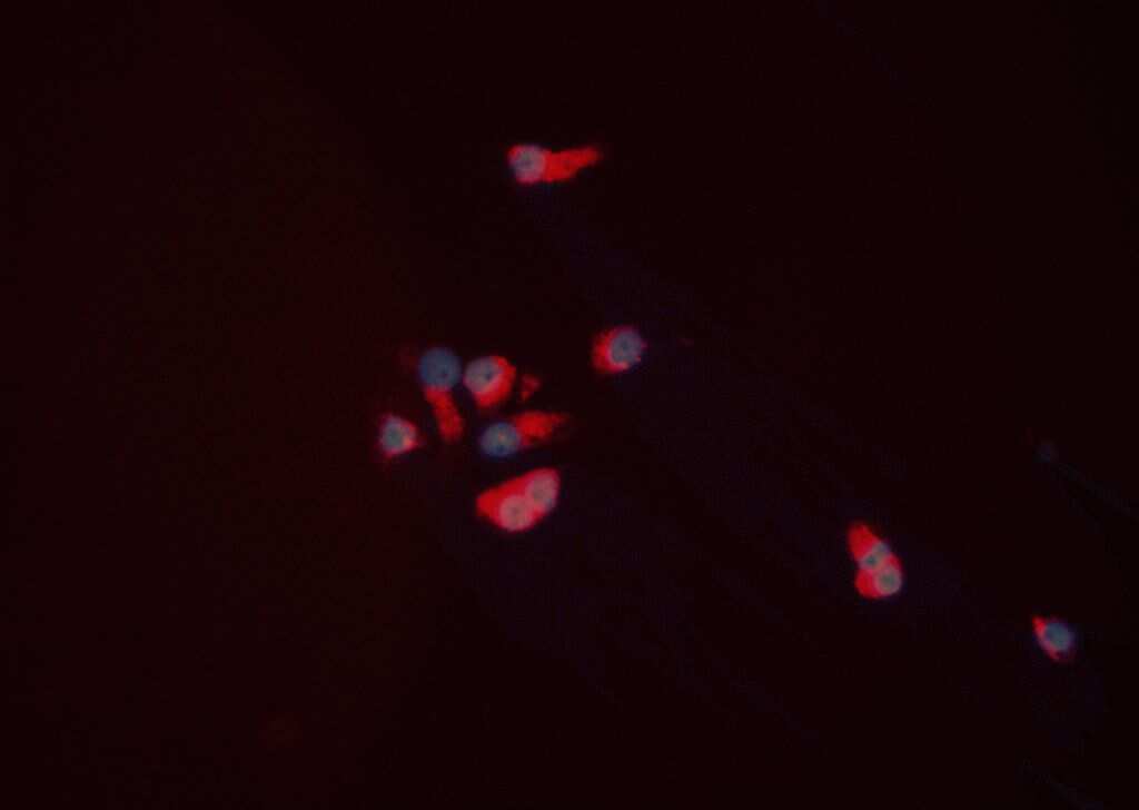
- Immunofluorescent analysis of NPY5R in HEPG2 cells. Samples were fixed with paraformaldehyde, permeabilized with 0.1% saponin, blocked with 10% serum (45 min at 25°C) incubated with NPY5R polyclonal antibody (Product # PA5-106850) using a dilution of 1:200 (1 hr, 37°C), and followed by goat anti-rabbit IgG Alexa Fluor 594 (red) at a dilution of 1:600.
- Submitted by
- Invitrogen Antibodies (provider)
- Main image
- Experimental details
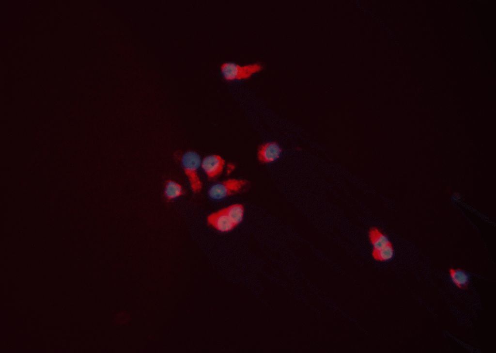
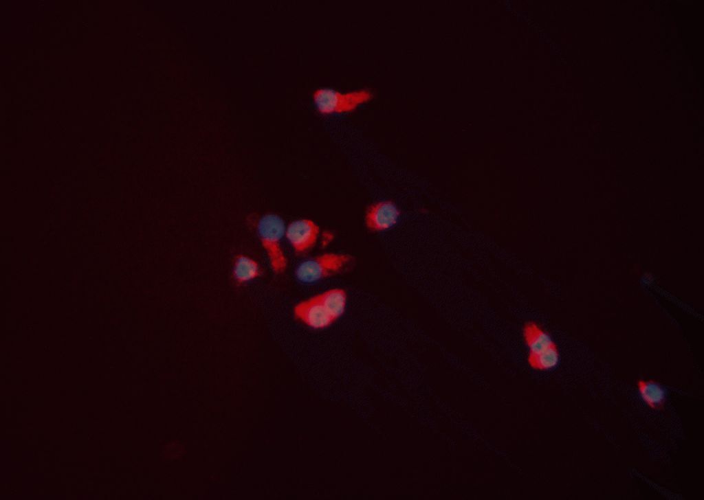
- Immunofluorescent analysis of NPY5R in HEPG2 cells. Samples were fixed with paraformaldehyde, permeabilized with 0.1% saponin, blocked with 10% serum (45 min at 25°C) incubated with NPY5R polyclonal antibody (Product # PA5-106850) using a dilution of 1:200 (1 hr, 37°C), and followed by goat anti-rabbit IgG Alexa Fluor 594 (red) at a dilution of 1:600.
- Submitted by
- Invitrogen Antibodies (provider)
- Main image
- Experimental details
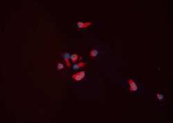
- Immunofluorescent analysis of NPY5R in HEPG2 cells. Samples were fixed with paraformaldehyde, permeabilized with 0.1% saponin, blocked with 10% serum (45 min at 25°C) incubated with NPY5R polyclonal antibody (Product # PA5-106850) using a dilution of 1:200 (1 hr, 37°C), and followed by goat anti-rabbit IgG Alexa Fluor 594 (red) at a dilution of 1:600.
Supportive validation
- Submitted by
- Invitrogen Antibodies (provider)
- Main image
- Experimental details
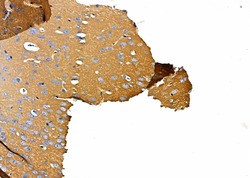
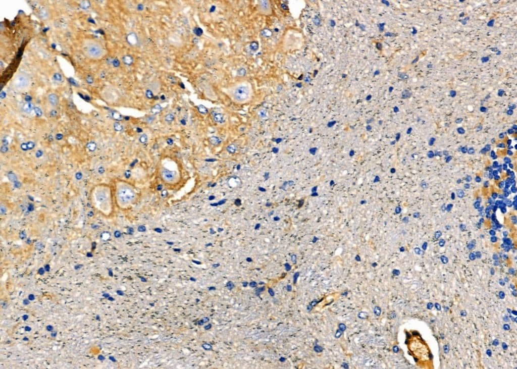
- Immunohistochemistry analysis of NPY5R in rat brain tissue. The sample was formaldehyde fixed and a heat mediated antigen retrieval step in citrate buffer was performed. Samples were incubated with NPY5R polyclonal antibody (Product # PA5-106850) using a dilution of 1:100 (4°C overnight) followed by HRP conjugated anti-Rabbit secondary antibody.

- Submitted by
- Invitrogen Antibodies (provider)
- Main image
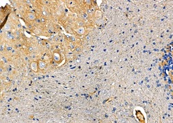
- Experimental details
- Immunohistochemistry analysis of NPY5R in mouse brain tissue. The sample was formaldehyde fixed and a heat mediated antigen retrieval step in citrate buffer was performed. Samples were incubated with NPY5R polyclonal antibody (Product # PA5-106850) using a dilution of 1:100 (4°C overnight) followed by HRP conjugated anti-Rabbit secondary antibody.
Supportive validation
- Submitted by
- Invitrogen Antibodies (provider)
- Main image

- Experimental details
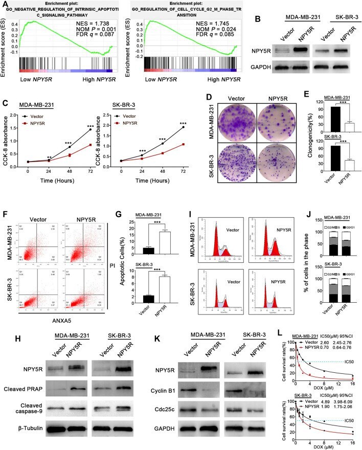
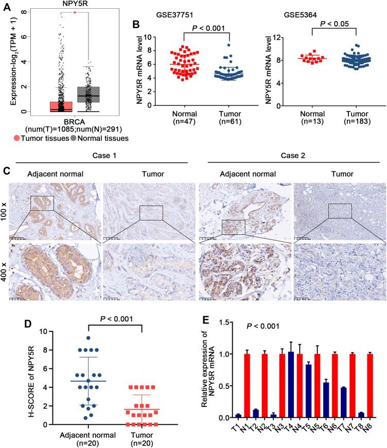
- FIGURE 2 The expression levels of NPY5R in BC tissues. (A) NPY5R mRNA expression in BC and normal breast tissues from TCGA database (* p < 0.05). (B) Analysis of NPY5R expression in BC and normal breast tissues using the GEO database. Statistical significance was evaluated using Wilcoxon rank sum test. (C) Immunohistochemical staining of NPY5R in BC tissues and adjacent non-tumor tissues. Typical images are shown at 200x and 400x magnifications. Scale bars, 50 mum. (D) H-SCORE of the two groups ( p < 0.001). (E) 8 pairs of primary BRCA and adjacent tissues are tested by q-PCR ( p < 0.001).
- Submitted by
- Invitrogen Antibodies (provider)
- Main image

- Experimental details
- FIGURE 5 Tumor suppressive functions of NPY5R in BC cells. (A) Gene enrichment plots showed that a series of gene sets including GO NEGATIVE REGULATION OF INTRINSIC APOPTOTIC SIGNALING PATHWAY (apoptosis) and GO REGULATION OF CELL CYCLE G2 M PHASE TRANSITION (cell cycle) were enriched in the NPY5R-high subgroup. (B) Overexpression of NPY5R in MDA-MB-231 and SK-BR-3 cells were confirmed by western blot. (C-E) The effects of transient NPY5R overexpression, the control vector, on cell proliferation and colony formation ability, as measured by CCK-8 (C) and colony formation (D,E) . Data represent the mean +- SD of three independent experiments; * p < 0.05; ** p < 0.01; *** p < 0.001. (F) The proportion of apoptotic cells in transiently transfected MDA-MB-231 and SK-BR-3 cells. (G) Quantification of apoptosis changes. (I,J) Flow cytometry analysis of cell cycle of transiently transfected MDA-MB-231 and SK-BR-3 cells by PI staining. (H,K) The expression of apoptosis-related proteins and cell cycle-related proteins in NPY5R-expressing cells was determined by western blot analysis. (L) CCK8 was performed to analyze effect of NPY5R expression on chemosensitivity of BC cells to DOX.