Explore
 Validate
 Learn
Learn Western blot
Antibody data
- Antibody Data
- Antigen structure
- References [0]
- Comments [0]
- Validations
- Western blot [2]
- Immunocytochemistry [2]
- Immunohistochemistry [1]
Submit
Validation data
Reference
Comment
Report error
- Product number
- AMR-024-25UL - Provider product page

- Provider
- Invitrogen Antibodies
- Product name
- MC4 Receptor (extracellular) Polyclonal Antibody
- Antibody type
- Polyclonal
- Antigen
- Other
- Reactivity
- Human, Mouse, Rat
- Host
- Rabbit
- Isotype
- IgG
- Vial size
- 25 µL
- Concentration
- 0.8 mg/mL
- Storage
- -20° C, Avoid Freeze/Thaw Cycles
No comments: Submit comment
Supportive validation
- Submitted by
- Invitrogen Antibodies (provider)
- Main image

- Experimental details
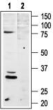
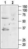
- Western blot analysis of rat brain lysates: - 1. Anti-MC4 Receptor (extracellular) Antibody (#AMR-024), (1:400). 2. Anti-MC4 Receptor (extracellular) Antibody , preincubated with MC4 Receptor (extracellular) Blocking Peptide (#BLP-MR024).
- Submitted by
- Invitrogen Antibodies (provider)
- Main image
- Experimental details
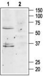
- Western blot analysis of mouse brain lysates: - 1. Anti-MC4 Receptor (extracellular) Antibody (#AMR-024), (1:400). 2. Anti-MC4 Receptor (extracellular) Antibody , preincubated with MC4 Receptor (extracellular) Blocking Peptide (#BLP-MR024).
Supportive validation
- Submitted by
- Invitrogen Antibodies (provider)
- Main image
- Experimental details
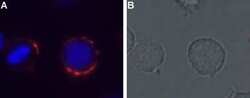
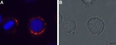
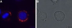
- Expression of MC4R inrat pituitary cell line - Cell surface detection of MC4R in live intact GH3 cells with Anti-MC4 Receptor (extracellular) Antibody (#AMR-024), (1:50). A. MC4R staining was observed,followed by Alexa-555-conjugated goat Anti-rabbit secondary Antibody (red).Hoechst 33342 (blue) is used to visualize the nuclei. B. Live view of the same field as A.
- Submitted by
- Invitrogen Antibodies (provider)
- Main image
- Experimental details
- Expression of MC4R inrat pituitary cell line - Cell surface detection of MC4R in live intact GH3 cells with Anti-MC4 Receptor (extracellular) Antibody (#AMR-024), (1:50). A. MC4R staining was observed,followed by Alexa-555-conjugated goat Anti-rabbit secondary Antibody (red).Hoechst 33342 (blue) is used to visualize the nuclei. B. Live view of the same field as A.

Supportive validation
- Submitted by
- Invitrogen Antibodies (provider)
- Main image

- Experimental details
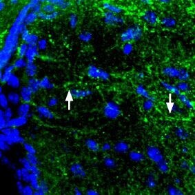
- Expression of MC4R in mouse brain - Immunohistochemical staining of perfusion-fixed frozen mouse brainsections using Anti-MC4 Receptor (extracellular) Antibody (#AMR-024), (1:100). MC4R (green)is expressedin the mouse hypothalamus in axonal processes (arrows). Hoechst 33342is used asthe counterstain (blue).