Explore
 Validate
 Learn
Learn Western blot
 Immunohistochemistry
Antibody data
- Antibody Data
- Antigen structure
- References [1]
- Comments [0]
- Validations
- Immunohistochemistry [1]
Submit
Validation data
Reference
Comment
Report error
- Product number
- PB9546 - Provider product page

- Provider
- Boster Biological Technology
- Product name
- Anti-CYP1B1 Antibody Picoband™
- Antibody type
- Polyclonal
- Description
- Polyclonal antibody for CYP1B1 detection. Host: Rabbit.Size: 100μg/vial. Tested applications: IHC-P. Reactive species: Human. CYP1B1 information: Molecular Weight: 60846 MW; Subcellular Localization: Endoplasmic reticulum membrane; Peripheral membrane protein. Microsome membrane; Peripheral membrane protein. Mitochondrion ; Tissue Specificity: Expressed in many tissues.
- Reactivity
- Human, Mouse, Rat
- Host
- Rabbit
- Vial size
- 100μg/vial
- Concentration
- Add 0.2ml of distilled water will yield a concentration of 500ug/ml.
- Storage
- At -20°C for one year. After reconstitution, at 4°C for one month. It can also be aliquoted and stored frozen at -20°C for a longer time. Avoid repeated freezing and thawing.
- Handling
- Add 0.2ml of distilled water will yield a concentration of 500ug/ml.
Submitted references Hepatic stellate cell stearoyl co-A desaturase activates leukotriene B4 receptor 2 - β-catenin cascade to promote liver tumorigenesis.
Sinha S, Aizawa S, Nakano Y, Rialdi A, Choi HY, Shrestha R, Pan SQ, Chen Y, Li M, Kapelanski-Lamoureux A, Yochum G, Sher L, Monga SP, Lazaris A, Machida K, Karin M, Guccione E, Tsukamoto H
Nature communications 2023 May 8;14(1):2651
Nature communications 2023 May 8;14(1):2651
No comments: Submit comment
Supportive validation
- Submitted by
- Boster Biological Technology (provider)
- Main image
- Experimental details
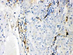
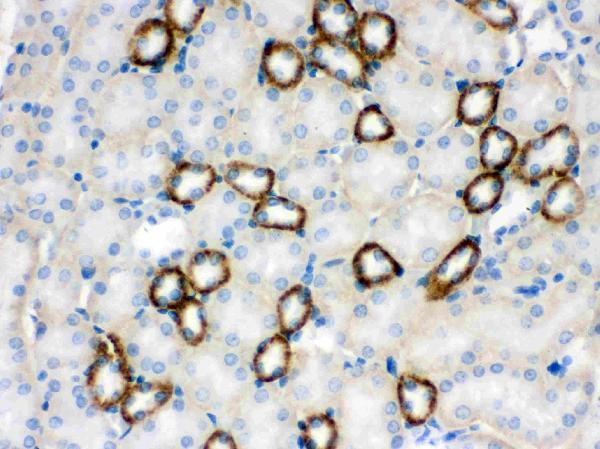
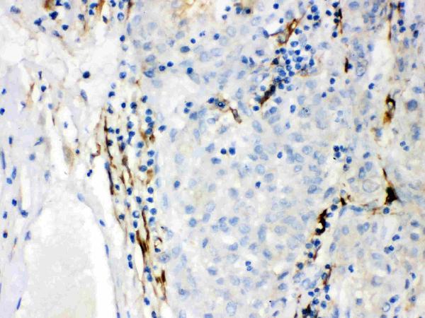
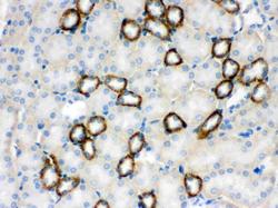
- IHC analysis of CYP1B1 using anti-CYP1B1 antibody (PB9546). CYP1B1 was detected in paraffin-embedded section of Mouse Kidney Tissue. Heat mediated antigen retrieval was performed in citrate buffer (pH6, epitope retrieval solution) for 20 mins. The tissue section was blocked with 10% goat serum. The tissue section was then incubated with 1μg/ml rabbit anti-CYP1B1 Antibody (PB9546) overnight at 4°C. Biotinylated goat anti-rabbit IgG was used as secondary antibody and incubated for 30 minutes at 37°C. The tissue section was developed using Strepavidin-Biotin-Complex (SABC)(Catalog # SA1022) with DAB as the chromogen.
- Additional image