Explore
 Validate
 Learn
Learn Western blot
 Immunocytochemistry
Antibody data
- Antibody Data
- Antigen structure
- References [0]
- Comments [0]
- Validations
- Immunocytochemistry [3]
- Immunoprecipitation [1]
- Flow cytometry [2]
Submit
Validation data
Reference
Comment
Report error
- Product number
- MA5-56469 - Provider product page

- Provider
- Invitrogen Antibodies
- Product name
- ADRM1 Recombinant Rabbit Monoclonal Antibody (PSH05-41)
- Antibody type
- Monoclonal
- Antigen
- Recombinant full-length protein
- Reactivity
- Human, Mouse, Rat
- Host
- Rabbit
- Isotype
- IgG
- Antibody clone number
- PSH05-41
- Vial size
- 100 μL
- Concentration
- 1 mg/mL
- Storage
- Store at 4°C short term. For long term storage, store at -20°C, avoiding freeze/thaw cycles.
No comments: Submit comment
Supportive validation
- Submitted by
- Invitrogen Antibodies (provider)
- Main image
- Experimental details
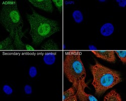
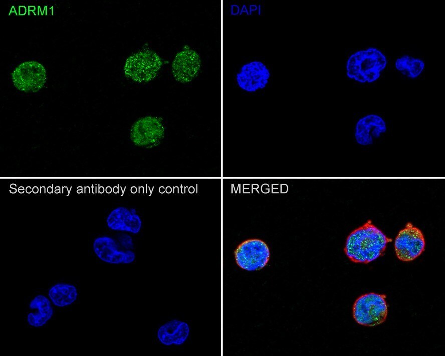
- Immunocytochemistry analysis of ADRM1 with SK-MEL-28 cells. Cells were fixed in 4% paraformaldehyde (20 min, room temp), permeabilized with 0.1% Triton X-100 in PBS (5 min, room temp), then blocked with 1% BSA in 10% negative goat serum (1 hr, room temp). Next they were incubated with ADRM1 Recombinant Monoclonal Antibody (Product # MA5-56469)at a dilution of 1:500 in 1% BSA in PBST overnight at 4 ℃. Followed by Goat Anti-Rabbit IgG H&L at a dilution of 1:1,000. DAPI was used to stain the cell nuclei (blue).

- Submitted by
- Invitrogen Antibodies (provider)
- Main image
- Experimental details
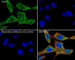
- Immunocytochemistry analysis of ADRM1 with NIH/3T3 cells. Cells were fixed in 4% paraformaldehyde (20 min, room temp), permeabilized with 0.1% Triton X-100 in PBS (5 min, room temp), then blocked with 1% BSA in 10% negative goat serum (1 hr, room temp). Next they were incubated with ADRM1 Recombinant Monoclonal Antibody (Product # MA5-56469)at a dilution of 1:500 in 1% BSA in PBST overnight at 4 ℃. Followed by Goat Anti-Rabbit IgG H&L at a dilution of 1:1,000. DAPI was used to stain the cell nuclei (blue).

- Submitted by
- Invitrogen Antibodies (provider)
- Main image
- Experimental details
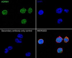
- Immunocytochemistry analysis of ADRM1 with PC-12 cells. Cells were fixed in 4% paraformaldehyde (20 min, room temp), permeabilized with 0.1% Triton X-100 in PBS (5 min, room temp), then blocked with 1% BSA in 10% negative goat serum (1 hr, room temp). Next they were incubated with ADRM1 Recombinant Monoclonal Antibody (Product # MA5-56469)at a dilution of 1:500 in 1% BSA in PBST overnight at 4 ℃. Followed by Goat Anti-Rabbit IgG H&L at a dilution of 1:1,000. DAPI was used to stain the cell nuclei (blue).
Supportive validation
- Submitted by
- Invitrogen Antibodies (provider)
- Main image
- Experimental details
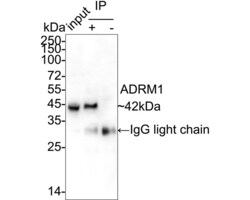
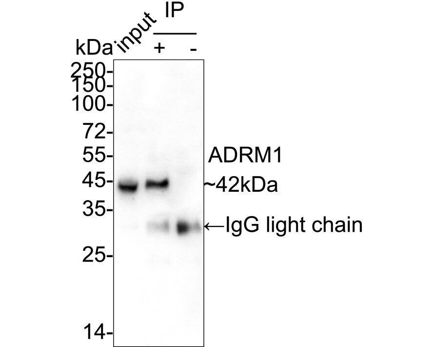
- ADRM1 was immunoprecipitated from 0.2 mg HEK-293 cell lysate with ADRM1 Recombinant Monoclonal Antibody (Product # MA5-56469) at 2 µg/25 µl agarose. Western blot was performed from the immunoprecipitate using ADRM1 Recombinant Monoclonal Antibody (Product # MA5-56469) at 1:1,000 dilution. Followed by Anti-Rabbit IgG for IP Nano-secondary antibody at 1:5,000 dilution (1 hr, room temperature). Lane 1: HEK-293 cell lysate; Lane 2: (MA5-56469) IP in HEK-293 cell lysate; Lane 3: Rabbit IgG in HEK-293 cell lysate. Blocking/Dilution buffer: 5% NFDM/TBST. Exposure time: 3 minutes.
Supportive validation
- Submitted by
- Invitrogen Antibodies (provider)
- Main image
- Experimental details
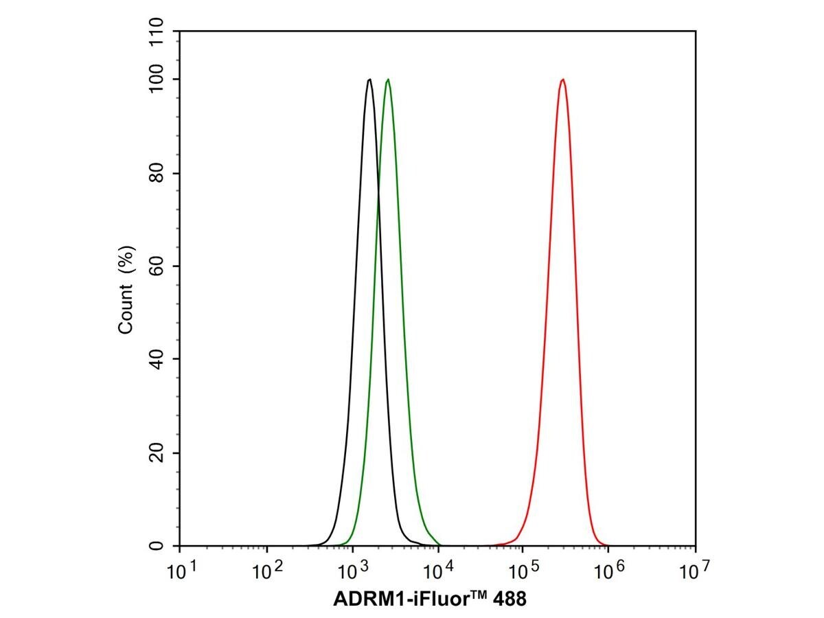
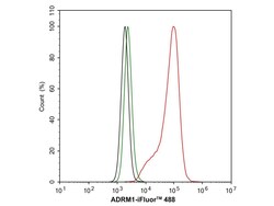
- Flow cytometry analysis of ADRM1 with PC-12 cells. The cells were fixed, permeabilized and incubated with ADRM1 Recombinant Monoclonal Antibody (Product # MA5-56469) at a dilution of 1:1,000 (red) and compared with Rabbit IgG Isotype Control (green). Followed by iFluor™ 488 conjugate-Goat anti-Rabbit IgG Secondary antibody at 1:1,000 dilution (30 min, 4℃).
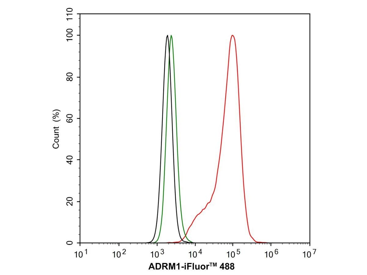
- Submitted by
- Invitrogen Antibodies (provider)
- Main image
- Experimental details
- Flow cytometry analysis of ADRM1 with NIH/3T3 cells. The cells were fixed, permeabilized and incubated with ADRM1 Recombinant Monoclonal Antibody (Product # MA5-56469) at a dilution of 1:1,000 (red) and compared with Rabbit IgG Isotype Control (green). Followed by iFluor™ 488 conjugate-Goat anti-Rabbit IgG Secondary antibody at 1:1,000 dilution (30 min, 4℃).