Explore
 Validate
 Learn
Learn Western blot
 Immunocytochemistry
 Immunoprecipitation
Antibody data
- Antibody Data
- Antigen structure
- References [6]
- Comments [0]
- Validations
- Immunocytochemistry [4]
- Immunohistochemistry [3]
Submit
Validation data
Reference
Comment
Report error
- Product number
- PA1-1741 - Provider product page

- Provider
- Invitrogen Antibodies
- Product name
- MUSK Polyclonal Antibody
- Antibody type
- Polyclonal
- Antigen
- Recombinant full-length protein
- Description
- PA1-1741 detects a predominant band at ~98kD. Other lower MW nonspecific bands of unknown identity were also detected and are likely to be proteolytic fragments.
- Reactivity
- Human, Mouse, Rat
- Host
- Rabbit
- Isotype
- IgG
- Vial size
- 200 μg
- Concentration
- 1 mg/mL
- Storage
- -20°C, Avoid Freeze/Thaw Cycles
Submitted references A 3D culture model of innervated human skeletal muscle enables studies of the adult neuromuscular junction.
Interaction between ROR1 and MuSK activation complex in myogenic cells.
The Agrin/MuSK signaling pathway is spatially segregated from the neuregulin/ErbB receptor signaling pathway at the neuromuscular junction.
Dimerization of the muscle-specific kinase induces tyrosine phosphorylation of acetylcholine receptors and their aggregation on the surface of myotubes.
Laminin and alpha-dystroglycan mediate acetylcholine receptor aggregation via a MuSK-independent pathway.
Tyrosine phosphorylation of the muscle-specific kinase is exclusively induced by acetylcholine receptor-aggregating agrin fragments.
Afshar Bakooshli M, Lippmann ES, Mulcahy B, Iyer N, Nguyen CT, Tung K, Stewart BA, van den Dorpel H, Fuehrmann T, Shoichet M, Bigot A, Pegoraro E, Ahn H, Ginsberg H, Zhen M, Ashton RS, Gilbert PM
eLife 2019 May 14;8
eLife 2019 May 14;8
Interaction between ROR1 and MuSK activation complex in myogenic cells.
Karvonen H, Summala K, Niininen W, Barker HR, Ungureanu D
FEBS letters 2018 Feb;592(3):434-445
FEBS letters 2018 Feb;592(3):434-445
The Agrin/MuSK signaling pathway is spatially segregated from the neuregulin/ErbB receptor signaling pathway at the neuromuscular junction.
Trinidad JC, Fischbach GD, Cohen JB
The Journal of neuroscience : the official journal of the Society for Neuroscience 2000 Dec 1;20(23):8762-70
The Journal of neuroscience : the official journal of the Society for Neuroscience 2000 Dec 1;20(23):8762-70
Dimerization of the muscle-specific kinase induces tyrosine phosphorylation of acetylcholine receptors and their aggregation on the surface of myotubes.
Hopf C, Hoch W
The Journal of biological chemistry 1998 Mar 13;273(11):6467-73
The Journal of biological chemistry 1998 Mar 13;273(11):6467-73
Laminin and alpha-dystroglycan mediate acetylcholine receptor aggregation via a MuSK-independent pathway.
Montanaro F, Gee SH, Jacobson C, Lindenbaum MH, Froehner SC, Carbonetto S
The Journal of neuroscience : the official journal of the Society for Neuroscience 1998 Feb 15;18(4):1250-60
The Journal of neuroscience : the official journal of the Society for Neuroscience 1998 Feb 15;18(4):1250-60
Tyrosine phosphorylation of the muscle-specific kinase is exclusively induced by acetylcholine receptor-aggregating agrin fragments.
Hopf C, Hoch W
European journal of biochemistry 1998 Apr 15;253(2):382-9
European journal of biochemistry 1998 Apr 15;253(2):382-9
No comments: Submit comment
Supportive validation
- Submitted by
- Invitrogen Antibodies (provider)
- Main image
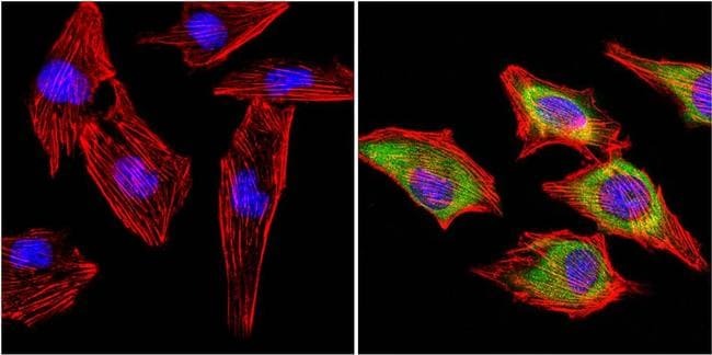
- Experimental details
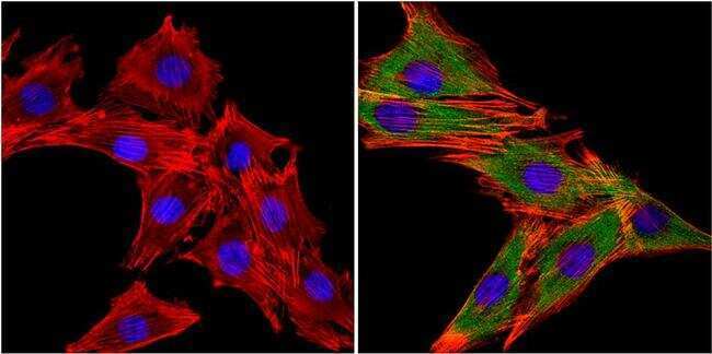
- Immunofluorescent analysis of muscle-specific kinase (MuSK) (green) in L6 rat myoblast cells. The cells were fixed with 4% paraformaldehyde for 15 minutes, permeabilized with 0.1% Triton X-100 in TBS for 10 minutes, and blocked with 3% BSA-PBS for 30 minutes at room temperature. Cells were stained without (left panel) or with a MuSK polyclonal antibody (Product # PA1-1741, right panel), at a dilution of 1:20 in blocking buffer for at least 1 hour at room temperature, and then incubated with a DyLight 488 goat anti-rabbit IgG secondary antibody (Product # 35552) for 45 minutes at room temperature (green). F-Actin (both panels, red) was stained with DyLight 554 Phalloidin (Product # 21834) and nuclei (both panels, blue) were stained with DAPI. Images were taken at 60X magnification.
- Submitted by
- Invitrogen Antibodies (provider)
- Main image
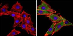
- Experimental details
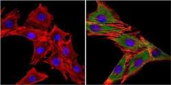
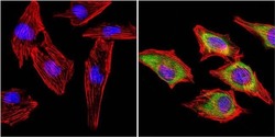
- Immunofluorescent analysis of muscle-specific kinase (MuSK) (green) in C2C12 mouse myoblast cells. The cells were fixed with 4% paraformaldehyde for 15 minutes, permeabilized with 0.1% Triton X-100 in TBS for 10 minutes, and blocked with 3% BSA-PBS for 30 minutes at room temperature. Cells were stained without (left panel) or with a MuSK polyclonal antibody (Product # PA1-1741, right panel), at a dilution of 1:20 in blocking buffer for at least 1 hour at room temperature, and then incubated with a DyLight 488 goat anti-rabbit IgG secondary antibody (Product # 35552) for 45 minutes at room temperature (green). F-Actin (both panels, red) was stained with DyLight 554 Phalloidin (Product # 21834) and nuclei (both panels, blue) were stained with DAPI. Images were taken at 60X magnification.
- Submitted by
- Invitrogen Antibodies (provider)
- Main image
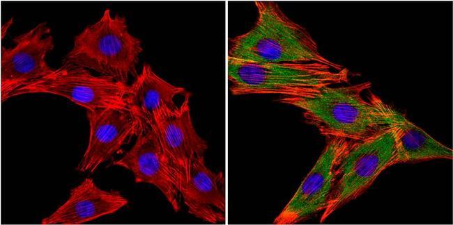
- Experimental details
- Immunofluorescent analysis of muscle-specific kinase (MuSK) (green) in L6 rat myoblast cells. The cells were fixed with 4% paraformaldehyde for 15 minutes, permeabilized with 0.1% Triton X-100 in TBS for 10 minutes, and blocked with 3% BSA-PBS for 30 minutes at room temperature. Cells were stained without (left panel) or with a MuSK polyclonal antibody (Product # PA1-1741, right panel), at a dilution of 1:20 in blocking buffer for at least 1 hour at room temperature, and then incubated with a DyLight 488 goat anti-rabbit IgG secondary antibody (Product # 35552) for 45 minutes at room temperature (green). F-Actin (both panels, red) was stained with DyLight 554 Phalloidin (Product # 21834) and nuclei (both panels, blue) were stained with DAPI. Images were taken at 60X magnification.
- Submitted by
- Invitrogen Antibodies (provider)
- Main image

- Experimental details
- Immunofluorescent analysis of muscle-specific kinase (MuSK) (green) in C2C12 mouse myoblast cells. The cells were fixed with 4% paraformaldehyde for 15 minutes, permeabilized with 0.1% Triton X-100 in TBS for 10 minutes, and blocked with 3% BSA-PBS for 30 minutes at room temperature. Cells were stained without (left panel) or with a MuSK polyclonal antibody (Product # PA1-1741, right panel), at a dilution of 1:20 in blocking buffer for at least 1 hour at room temperature, and then incubated with a DyLight 488 goat anti-rabbit IgG secondary antibody (Product # 35552) for 45 minutes at room temperature (green). F-Actin (both panels, red) was stained with DyLight 554 Phalloidin (Product # 21834) and nuclei (both panels, blue) were stained with DAPI. Images were taken at 60X magnification.

Supportive validation
- Submitted by
- Invitrogen Antibodies (provider)
- Main image
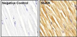
- Experimental details
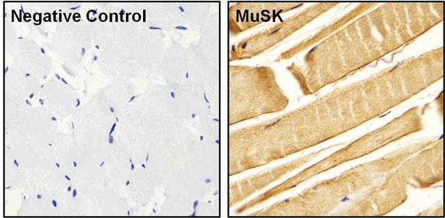
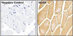
- Immunohistochemistry was performed on rat skeletal muscle tissue. To expose target proteins, heat induced antigen retrieval was performed using 10mM sodium citrate (pH 6.0) buffer for 10 minutes using a microwave. Following antigen retrieval, tissues were blocked in 3% BSA-PBS for 30 minutes and then probed with (right panel) or without (left panel) a MuSK polyclonal antibody (Product # PA1-1741) at a dilution of 1:20 overnight at 4C in a humidified chamber. Tissues were washed extensively with TBS + 0.025% Triton X-100 (Product # 28314) and endogenous peroxidase activity quenched with Peroxidase Suppressor (Product # 35000) for 30 minutes at room temperature. Detection was performed using a goat anti-rabbit HRP secondary antibody (Product # 31460) followed by colorimetric detection using metal enhanced DAB (Product # 34065). Tissues were counterstained with hematoxylin and prepped for mounting.

- Submitted by
- Invitrogen Antibodies (provider)
- Main image
- Experimental details
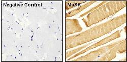
- Immunohistochemistry was performed on mouse skeletal muscle tissue. To expose target proteins, heat induced antigen retrieval was performed using 10mM sodium citrate (pH 6.0) buffer for 10 minutes using a microwave. Following antigen retrieval, tissues were blocked in 3% BSA-PBS for 30 minutes and then probed with (right panel) or without (left panel) a MuSK polyclonal antibody (Product # PA1-1741) at a dilution of 1:20 overnight at 4C in a humidified chamber. Tissues were washed extensively with TBS + 0.025% Triton X-100 (Product # 28314) and endogenous peroxidase activity quenched with Peroxidase Suppressor (Product # 35000) for 30 minutes at room temperature. Detection was performed using a goat anti-rabbit HRP secondary antibody (Product # 31460) followed by colorimetric detection using metal enhanced DAB (Product # 34065). Tissues were counterstained with hematoxylin and prepped for mounting.

- Submitted by
- Invitrogen Antibodies (provider)
- Main image
- Experimental details
- Immunohistochemistry was performed on human skeletal muscle tissue. To expose target proteins, heat induced antigen retrieval was performed using 10mM sodium citrate (pH 6.0) buffer for 10 minutes using a microwave. Following antigen retrieval, tissues were blocked in 3% BSA-PBS for 30 minutes and then probed with (right panel) or without (left panel) a MuSK polyclonal antibody (Product # PA1-1741) at a dilution of 1:20 overnight at 4C in a humidified chamber. Tissues were washed extensively with TBS + 0.025% Triton X-100 (Product # 28314) and endogenous peroxidase activity quenched with Peroxidase Suppressor (Product # 35000) for 30 minutes at room temperature. Detection was performed using a goat anti-rabbit HRP secondary antibody (Product # 31460) followed by colorimetric detection using metal enhanced DAB (Product # 34065). Tissues were counterstained with hematoxylin and prepped for mounting.