Explore
 Validate
 Learn
Learn Western blot
 Immunocytochemistry
Antibody data
- Antibody Data
- Antigen structure
- References [3]
- Comments [0]
- Validations
- Immunocytochemistry [1]
- Other assay [2]
Submit
Validation data
Reference
Comment
Report error
- Product number
- 702516 - Provider product page

- Provider
- Invitrogen Antibodies
- Product name
- P2Y12 Recombinant Rabbit Monoclonal Antibody (4H5L19)
- Antibody type
- Monoclonal
- Antigen
- Synthetic peptide
- Description
- This antibody is predicted to react with Monkey, Cat, Dog Recombinant rabbit monoclonal antibodies are produced using in vitro expression systems. The expression systems are developed by cloning in the specific antibody DNA sequences from immunoreactive rabbits. Then, individual clones are screened to select the best candidates for production. The advantages of using recombinant rabbit monoclonal antibodies include: better specificity and sensitivity, lot-to-lot consistency, animal origin-free formulations, and broader immunoreactivity to diverse targets due to larger rabbit immune repertoire.
- Reactivity
- Human, Mouse, Rat
- Host
- Rabbit
- Isotype
- IgG
- Antibody clone number
- 4H5L19
- Vial size
- 100 μg
- Concentration
- 0.5 mg/mL
- Storage
- Store at 4°C short term. For long term storage, store at -20°C, avoiding freeze/thaw cycles.
Submitted references Crosstalk between astrocytes and microglia results in increased degradation of α-synuclein and amyloid-β aggregates.
Inhibiting P2Y12 in Macrophages Induces Endoplasmic Reticulum Stress and Promotes an Anti-Tumoral Phenotype.
Elevated protein synthesis in microglia causes autism-like synaptic and behavioral aberrations.
Rostami J, Mothes T, Kolahdouzan M, Eriksson O, Moslem M, Bergström J, Ingelsson M, O'Callaghan P, Healy LM, Falk A, Erlandsson A
Journal of neuroinflammation 2021 Jun 3;18(1):124
Journal of neuroinflammation 2021 Jun 3;18(1):124
Inhibiting P2Y12 in Macrophages Induces Endoplasmic Reticulum Stress and Promotes an Anti-Tumoral Phenotype.
Pavlović N, Kopsida M, Gerwins P, Heindryckx F
International journal of molecular sciences 2020 Oct 31;21(21)
International journal of molecular sciences 2020 Oct 31;21(21)
Elevated protein synthesis in microglia causes autism-like synaptic and behavioral aberrations.
Xu ZX, Kim GH, Tan JW, Riso AE, Sun Y, Xu EY, Liao GY, Xu H, Lee SH, Do NY, Lee CH, Clipperton-Allen AE, Kwon S, Page DT, Lee KJ, Xu B
Nature communications 2020 Apr 14;11(1):1797
Nature communications 2020 Apr 14;11(1):1797
No comments: Submit comment
Supportive validation
- Submitted by
- Invitrogen Antibodies (provider)
- Main image
- Experimental details
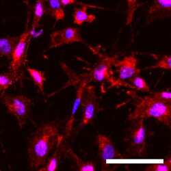
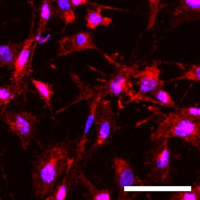
- Immunofluorescent analysis of P2Y12 (red) in human iPSC-derived microglia. The cells were fixed with 4% paraformaldehyde for 15 min at room temperature, and then permeabilized and blocked with 0.25% Triton X-100 and 10% donkey serum in PBS for 20 min. Samples were then incubated with Anti-P2Y12 Recombinant Rabbit Monoclonal Antibody (red; Product # 702516) at a dilution of 1:500 in PBS containing 0.25% Triton X-100 and 10% donkey serum at 48304C overnight, followed by incubation with Donkey Anti-Rabbit Alexa Fluor 568 (Product # A10042) at a dilution of 1:1000 as well as DAPI (blue; 1:25000) in PBS containing 0.25% Triton X-100 and 10% donkey serum at room temperature for 1 hour. Images were taken at 20X magnification. (Scale bar: 50 µm) Data courtesy of Dr. Zhexing Wen at Emory University.
Supportive validation
- Submitted by
- Invitrogen Antibodies (provider)
- Main image
- Experimental details
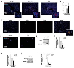
- Figure 1 The P2Y12 receptor is expressed in mouse models for liver cirrhosis and hepatocellular carcinoma. ( A ) Immunohistochemical staining with P2Y12 antibodies of liver sections from healthy mice, mice with carbon tetrachloride (CCL 4) -induced liver cirrhosis and mice with diethylnitrosamine (DEN)-induced liver cancer. ( B ) Quantification of immunohistochemical staining with P2Y12 antibodies. ( C ) Co-staining of P2Y12 and macrophage marker F4/80 in liver slides from DEN-induced hepatocellular carcinoma (HCC). ( D ) Immunocytochemical staining of THP1-differentiated macrophages and Raw264.7 cells. ( E ) Western Blot image of P2Y12 protein expression and beta-actin protein in THP1-differentiated macrophages and Raw264.7 cells. ( F ) mRNA expression levels of P2yr12 in interferon gamma and lipopolysaccharide-stimulated anti-tumoral (M1) macrophages and interleukin (IL)-13 and IL-4-stimulated pro-tumoral (M2) macrophages ( G ) Quantification of protein expression in pro-tumoral (M2) and anti-tumoral (M1) macrophages. ( H ) Representative image of Western blot showing P2Y12 protein expression in pro-tumoral (M2) and anti-tumoral (M1) macrophages. ( I ) mRNA expression of P2yr12 in livers from healthy mice, mice with CCL4-induced liver cirrhosis and mice with DEN-induced liver cancer. * = p < 0.05; ** = p < 0.01.

- Submitted by
- Invitrogen Antibodies (provider)
- Main image
- Experimental details
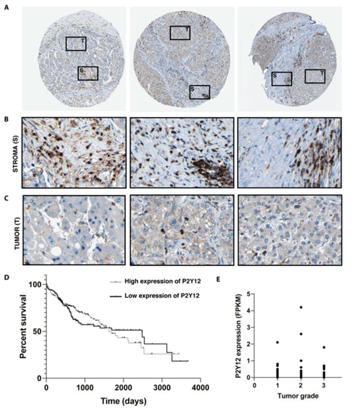
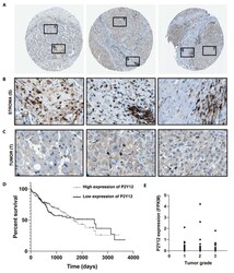
- Figure 2 Expression of P2Y12 is located in the stroma of patients with hepatocellular carcinoma. ( A ) Micrographs of hepatocellular carcinoma biopsies stained with P2Y12 antibodies, obtained from the Human Protein Atlas, marking tumor (T) and stomal (S) area [,]. ( B ) Detail of stroma and ( C ) tumor areas within the biopsies []. ( D ) Kaplan-Meier survival curve of HCC patients with high or low expression of P2Y12 obtained through the Human Protein Atlas []. ( E ) P2Y12 expression in different HCC stages, measured as number fragments per kilobase of exon per million reads (RPKM) from the Human Protein Atlas [].