 Explore
 Validate
 Learn
Learn Western blot
 Immunohistochemistry
Antibody data
- Antibody Data
- Antigen structure
- References [2]
- Comments [0]
- Validations
- Immunohistochemistry [3]
- Other assay [2]
Submit
Validation data
Reference
Comment
Report error
- Product number
- PA5-42106 - Provider product page

- Provider
- Invitrogen Antibodies
- Product name
- TGM4 Polyclonal Antibody
- Antibody type
- Polyclonal
- Antigen
- Synthetic peptide
- Description
- Peptide sequence: KEDMVFMPDE DERKEYILND TGCHYVGAAR SIKCKPWNFG QFEKNVLDCC Sequence homology: Guinea Pig: 75%; Human: 100%; Mouse: 83%; Rat: 75%; Zebrafish: 82%
- Reactivity
- Human
- Host
- Rabbit
- Isotype
- IgG
- Vial size
- 100 μL
- Concentration
- 0.5 mg/mL
- Storage
- -20°C, Avoid Freeze/Thaw Cycles
Submitted references Comprehensive Proteomic Profiling of Urinary Exosomes and Identification of Potential Non-invasive Early Biomarkers of Alzheimer's Disease in 5XFAD Mouse Model.
A single-cell atlas of the mouse and human prostate reveals heterogeneity and conservation of epithelial progenitors.
Song Z, Xu Y, Zhang L, Zhou L, Zhang Y, Han Y, Li X, Yu P, Qu Y, Zhao W, Qin C
Frontiers in genetics 2020;11:565479
Frontiers in genetics 2020;11:565479
A single-cell atlas of the mouse and human prostate reveals heterogeneity and conservation of epithelial progenitors.
Crowley L, Cambuli F, Aparicio L, Shibata M, Robinson BD, Xuan S, Li W, Hibshoosh H, Loda M, Rabadan R, Shen MM
eLife 2020 Sep 11;9
eLife 2020 Sep 11;9
No comments: Submit comment
Supportive validation
- Submitted by
- Invitrogen Antibodies (provider)
- Main image
- Experimental details
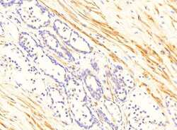
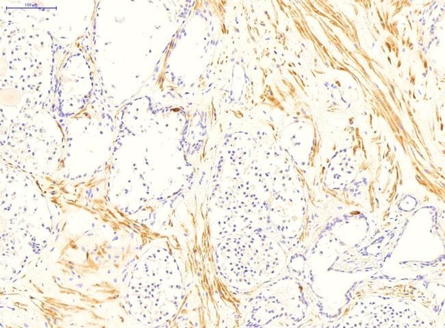
- Immunohistochemistry analysis of normal human prostate cells using an anti-Transglutaminase 4 polyclonal antibody (Product # PA5-42106).

- Submitted by
- Invitrogen Antibodies (provider)
- Main image
- Experimental details
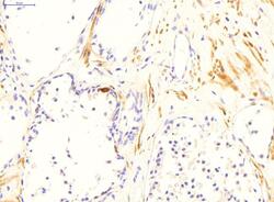
- Immunohistochemistry analysis of normal human prostate cells using an anti-Transglutaminase 4 polyclonal antibody (Product # PA5-42106). Primary Antibody Dilution: 2 µg/mL.
- Submitted by
- Invitrogen Antibodies (provider)
- Main image

- Experimental details
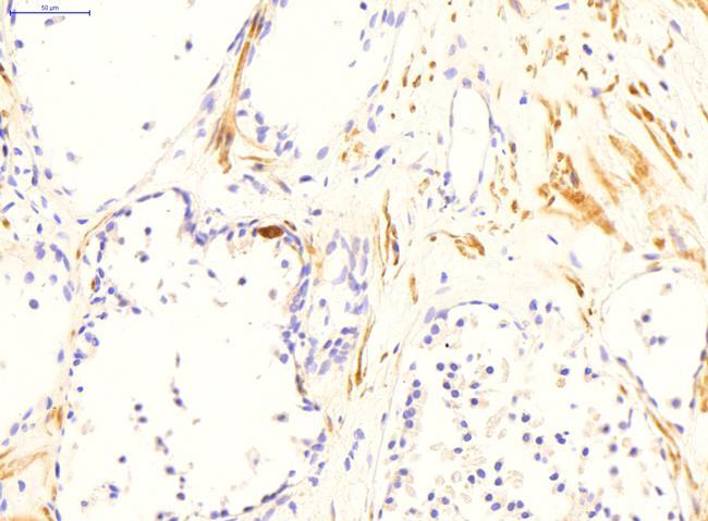
- Immunohistochemistry analysis of normal human prostate cells using an anti-Transglutaminase 4 polyclonal antibody (Product # PA5-42106). Primary Antibody Dilution: 2 µg/mL.
Supportive validation
- Submitted by
- Invitrogen Antibodies (provider)
- Main image
- Experimental details
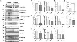
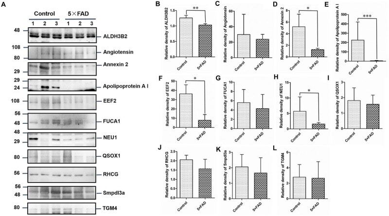
- FIGURE 7 Western blots (WB) and quantitative analysis of the other 11 differential proteins in the urinary exosomes isolated from control and 5XFAD mice. (A) Same aliquot of urinary exosomes isolated from three control samples and three 5XFAD mice samples. ALDH3B2, Angiotensin, Annexin 2, Apolipoprotein A I, EEF2, FUCA1, NEU1, QSOX1, RHCG, Smpdl3a, and TGM4 proteins were detected by WB in sequence. Representative blots of the tested proteins and the molecular weight of standard proteins were presented. (B-L) Quantitative analysis of each of the tested protein's relative fold change, respectively. All data are presented as mean +- SD in triplicate experiments; Student's t -test; * P < 0.05; ** P < 0.01.
- Submitted by
- Invitrogen Antibodies (provider)
- Main image
- Experimental details
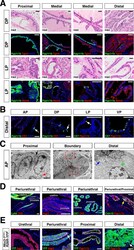
- Figure 2. Luminal epithelial populations display spatial and morphological heterogeneity. ( A ) H and E and IF of serial sections from the DP and LP, showing expression of proximal ( Ppp1r1b ) and distal ( Tgm4, Msmb ) markers; note apparent differences in the boundary regions of the two lobes. ( B ) Detection of distally localized LumP cells (arrows) in all four lobes; these are most abundant in the VP. ( C ) Scanning electron micrographs of the boundary region of the AP; central low-power image is flanked by high-power images of boxed regions. Red arrow, mitochondria; black arrow, membrane interdigitation; blue arrow, Golgi apparatus; green arrow, rough endoplasmic reticulum. ( D ) Identification of the periurethral region. Cells in the periurethral region generally express Ly6d, Ck7, Aqp3, and Ppp1r1b; notably, Cldn10-expressing LumP cells decrease approaching the periurethral region ( E ) Lineage-marking in Nkx3.1 Cre/+ ; R26R-YFP mice (n = 3) shows widespread YFP expression in the periurethral, proximal, and distal AP; small patches remain unrecombined and lack YFP (arrows). Scale bars in ( A,B,D,E ) indicate 50 um; scale bars in ( C ) indicate 2 um. Figure 2-figure supplement 1. Additional analysis of proximal-distal heterogeneity. ( A ) H and E and IF of serial sections from the AP and VP, showing expression of proximal ( Ppp1r1b ) and distal ( Tgm4, Trpv6 ) markers. ( B ) Scanning electron micrographs of proximal and distal regions of the AP. Left: red arrows, mitocho
