Explore
 Validate
 Learn
Learn Immunocytochemistry
Antibody data
- Antibody Data
- Antigen structure
- References [1]
- Comments [0]
- Validations
- Immunocytochemistry [2]
- Immunohistochemistry [1]
- Other assay [2]
Submit
Validation data
Reference
Comment
Report error
- Product number
- PA5-111083 - Provider product page

- Provider
- Invitrogen Antibodies
- Product name
- MAML1 Polyclonal Antibody
- Antibody type
- Polyclonal
- Antigen
- Recombinant protein fragment
- Description
- Immunogen sequence: QLGSPQVRAG SAGQTFLGPS SAPVSTDSPS LGGSQTLFHT SGQPRADNPS PNLMPASAQA QNAQRALAGV VLPSQGPGGA SELSSAHQLQ QIAAKQKREQ MLQNPQ
- Reactivity
- Human
- Host
- Rabbit
- Isotype
- IgG
- Vial size
- 100 μL
- Concentration
- 0.10 mg/mL
- Storage
- Store at 4°C short term. For long term storage, store at -20°C, avoiding freeze/thaw cycles.
Submitted references MAML1: a coregulator that alters endometrial epithelial cell adhesive capacity.
Zafir S, Zhou W, Menkhorst E, Santos L, Dimitriadis E
Fertility research and practice 2021 Mar 27;7(1):8
Fertility research and practice 2021 Mar 27;7(1):8
No comments: Submit comment
Supportive validation
- Submitted by
- Invitrogen Antibodies (provider)
- Main image
- Experimental details
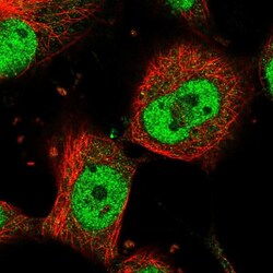
- Immunofluorescent staining of MAML1 in human cell line U-251 MG using MAML1 Polyclonal Antibody (Product # PA5-111083) shows localization to nucleoplasm.

- Submitted by
- Invitrogen Antibodies (provider)
- Main image

- Experimental details
- Immunofluorescent analysis of MAML1 in human cell line U-251 MG using MAML1 Polyclonal Antibody (Product # PA5-111083). Staining shows localization to nucleoplasm.

Supportive validation
- Submitted by
- Invitrogen Antibodies (provider)
- Main image
- Experimental details
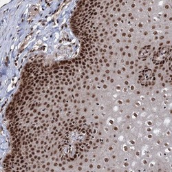
- Immunohistochemical analysis of MAML1 in human esophagus using MAML1 Polyclonal Antibody (Product # PA5-111083) shows strong nuclear positivity in squamous epithelial cells.

Supportive validation
- Submitted by
- Invitrogen Antibodies (provider)
- Main image
- Experimental details
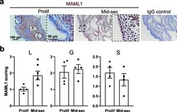
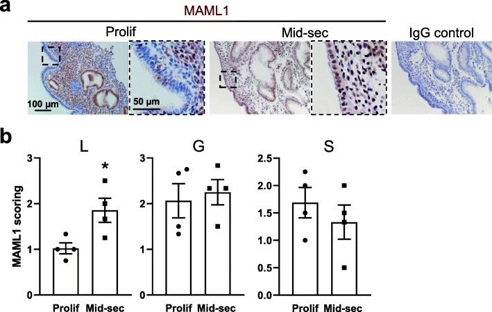
- Fig. 1 Comparison of MAML1 expression in fertile proliferative phase and mid-secretory phase endometrium. a Immunohistochemistry detection of MAML1 in luminal epithelium (L) glandular epithelium (G) and stroma (S) of human endometrium. A nuclear labeling was recorded in all cell types. The specificity of MAML1 labeling was confirmed through the inclusion of an isotype control in which the non-immune antibody of the same isotype was substituted for the MAML1 antibody at the same concertation. Sections were counterstained with hemotoxylin to highlight the cell nuclei (blue). b Staining intensity of MAML1 was semi-quantitated by scoring staining in tissues blinded to cycle stage. Data were presented as mean +- SEM. ( n = 4). * P < 0.05
- Submitted by
- Invitrogen Antibodies (provider)
- Main image

- Experimental details
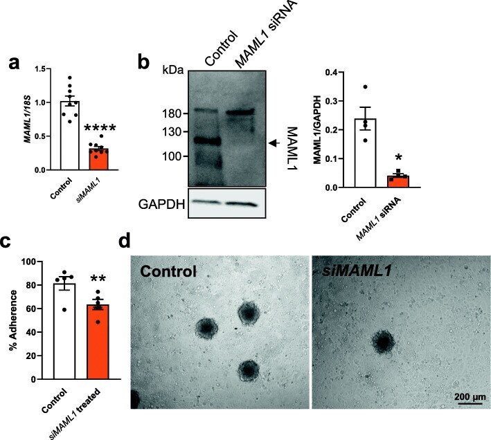
- Fig. 2 Examination of the effect of MAML1 knockdown on Ishikawa cell adhesive capacity. Ishikawa cells were transfected with either MAML1 siRNA (20 nM) or scrambled control (20 nM) before HTR8/SVneo spheroid adhesion assay or other analysis. a MAML1 knockdown was confirmed by qPCR. Expression levels were normalized to 18S ( n = 9). b MAML1 knockdown was confirmed by immunoblotting and densitometry, normalized against a loading control GAPDH ( n = 4). c MAML1 knockdown significantly compromised the spheroid adhesion compared to scrambled control ( n = 5). d Representative images are presented to show attached spheroids on the Ishikawa cell monolayer after adhesion assay. Data were presented as mean +- SEM. * P < 0.05, ** P < 0.01, *** P < 0.001