Explore
 Validate
 Learn
Learn Western blot
 Immunohistochemistry
Antibody data
- Antibody Data
- Antigen structure
- References [1]
- Comments [0]
- Validations
- Immunohistochemistry [7]
- Other assay [1]
Submit
Validation data
Reference
Comment
Report error
- Product number
- PA5-79399 - Provider product page

- Provider
- Invitrogen Antibodies
- Product name
- HSD11B2 Polyclonal Antibody
- Antibody type
- Polyclonal
- Antigen
- Synthetic peptide
- Description
- Reconstitute with 0.2 mL of distilled water to yield a concentration of 500 µg/mL. Positive Control - WB: rat kidney tissue, mouse kidney tissue, human placenta tissue. IHC: Mouse Pancreas tissue, Rat Pancreas tissue, Human Placenta tissue IHC-F: human placenta tissue, mouse kidney tissue, rat kidney tissue.
- Reactivity
- Human, Mouse, Rat
- Host
- Rabbit
- Isotype
- IgG
- Vial size
- 100 μg
- Concentration
- 500 μg/mL
- Storage
- -20°C
Submitted references Metabolomics analysis of follicular fluid coupled with oocyte aspiration reveals importance of glucocorticoids in primate periovulatory follicle competency.
Ravisankar S, Hanna CB, Brooks KE, Murphy MJ, Redmayne N, Ryu J, Kinchen JM, Chavez SL, Hennebold JD
Scientific reports 2021 Mar 22;11(1):6506
Scientific reports 2021 Mar 22;11(1):6506
No comments: Submit comment
Supportive validation
- Submitted by
- Invitrogen Antibodies (provider)
- Main image
- Experimental details
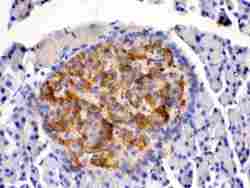
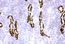
- Immunohistochemistry analysis of HSD11B2 on paraffin-embedded rat pancreas tissue. Sample was incubated with HSD11B2 polyclonal antibody (Product# PA5-79399).
- Submitted by
- Invitrogen Antibodies (provider)
- Main image
- Experimental details
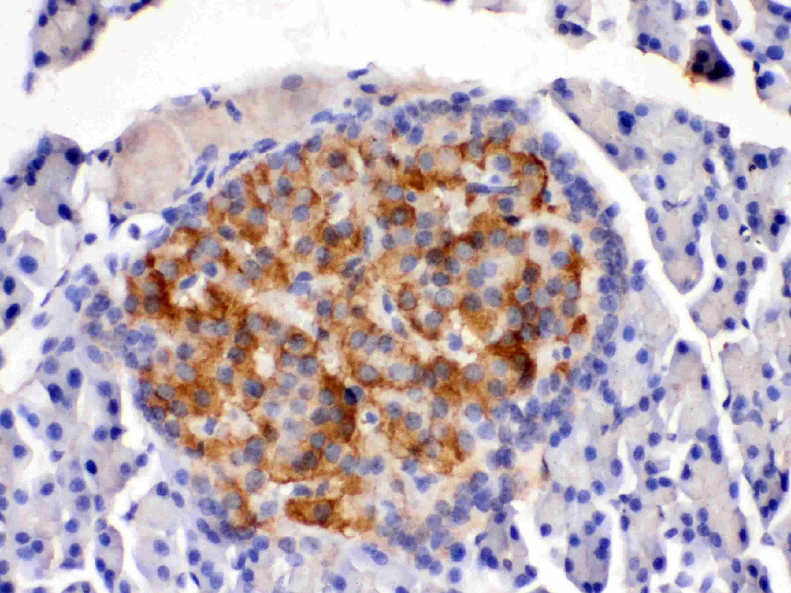
- Immunohistochemistry analysis of HSD11B2 on paraffin-embedded mouse pancreas tissue. Sample was incubated with HSD11B2 polyclonal antibody (Product# PA5-79399).
- Submitted by
- Invitrogen Antibodies (provider)
- Main image
- Experimental details
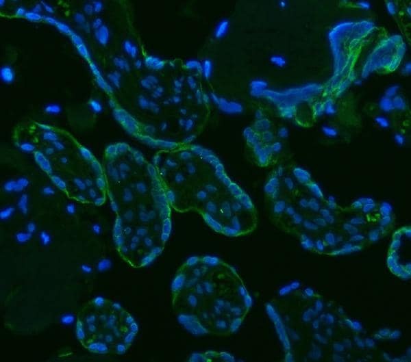
- Immunohistochemistry (Frozen) analysis of HSD11B2 in frozen section of human placenta tissue using HSD11B2 Polyclonal Antibody (Product # PA5-79399). The tissue section was blocked with 10% goat serum. The tissue section was then incubated with the primary antibody at 5 µg/mL overnight at 4°C. Peroxidase conjugated goat anti-rabbit IgG was used as secondary antibody and incubated for 30 minutes at 37°C. The tissue section was developed using HRP conjugated rabbit IgG Super Vision Assay Kit with DAB as the chromogen.

- Submitted by
- Invitrogen Antibodies (provider)
- Main image
- Experimental details
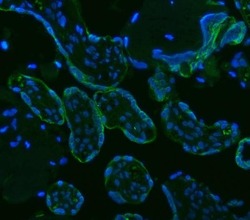
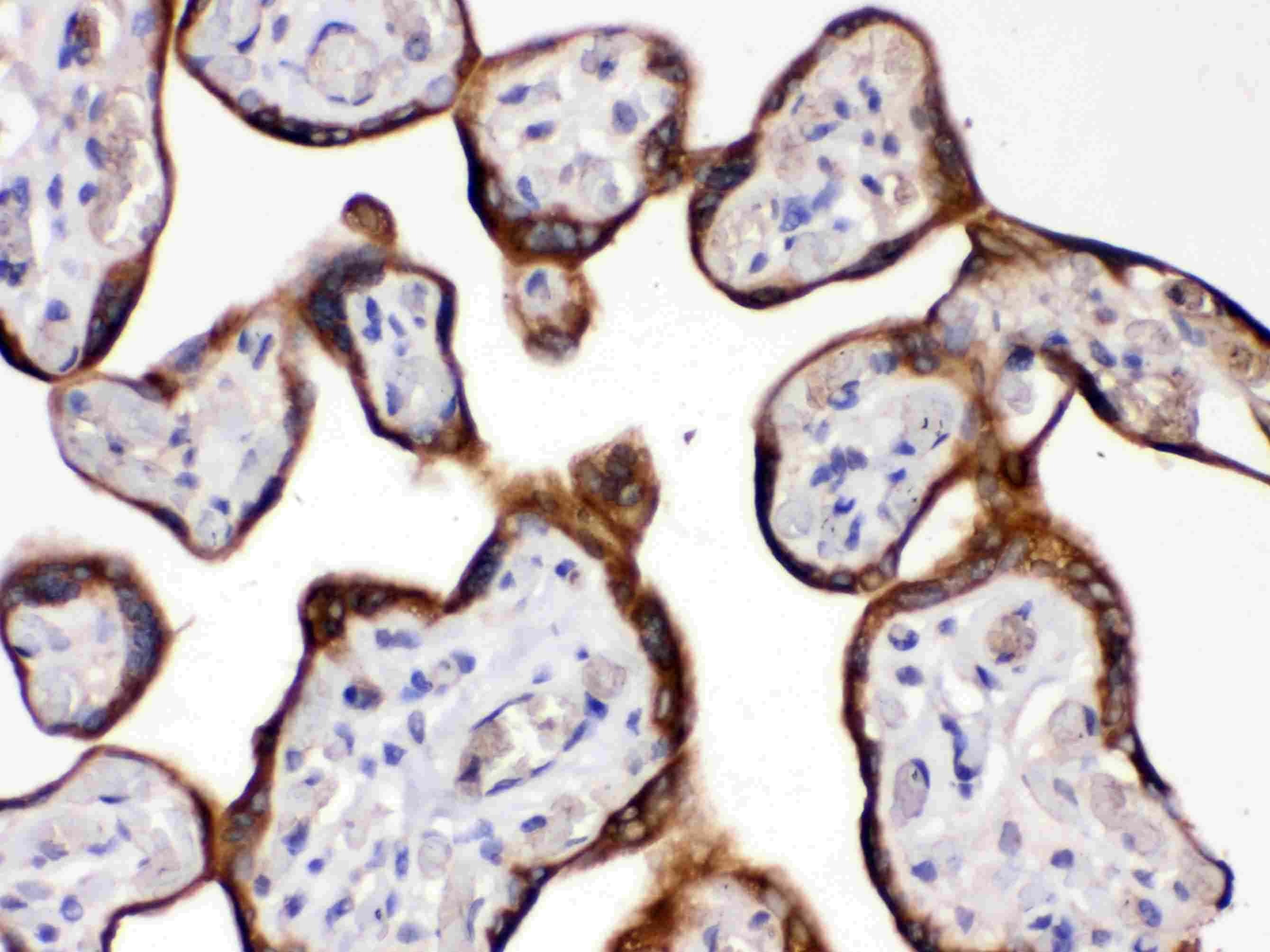
- Immunohistochemistry (Paraffin) analysis of HSD11B2 in paraffin-embedded section of human placenta tissue using HSD11B2 Polyclonal Antibody (Product # PA5-79399). Heat mediated antigen retrieval was performed in EDTA buffer (pH 8.0, epitope retrieval solution). The tissue section was blocked with 10% goat serum. The tissue section was then incubated with the primary antibody at a 5 µg/mL dilution overnight at 4°C. DyLight 488 conjugated goat anti-rabbit IgG was used as secondary antibody at 1:500 dilution and incubated for 30 minutes at 37°C. The section was counterstained with DAPI. Visualize using a fluorescence microscope and filter sets appropriate for the label used.
- Submitted by
- Invitrogen Antibodies (provider)
- Main image

- Experimental details
- Immunohistochemistry (Frozen) analysis of HSD11B2 in frozen section of mouse kidney tissue using HSD11B2 Polyclonal Antibody (Product # PA5-79399). The tissue section was blocked with 10% goat serum. The tissue section was then incubated with the primary antibody at a 2 µg/mL dilution overnight at 4°C. Peroxidase conjugated goat anti-rabbit IgG was used as secondary antibody and incubated for 30 minutes at 37°C. The tissue section was developed using HRP conjugated rabbit IgG Super Vision Assay Kit with DAB as the chromogen.
- Submitted by
- Invitrogen Antibodies (provider)
- Main image
- Experimental details
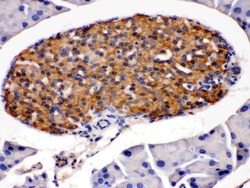
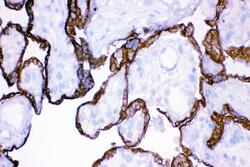
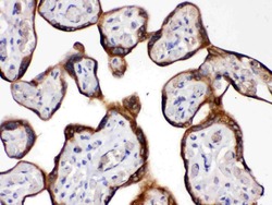
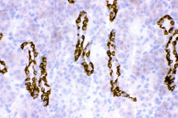
- Immunohistochemistry analysis of HSD11B2 on paraffin-embedded human placenta tissue. Sample was incubated with HSD11B2 polyclonal antibody (Product# PA5-79399).
- Submitted by
- Invitrogen Antibodies (provider)
- Main image
- Experimental details
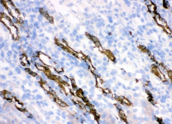
- Immunohistochemistry (Frozen) analysis of HSD11B2 in frozen section of rat kidney tissue using HSD11B2 Polyclonal Antibody (Product # PA5-79399). The tissue section was blocked with 10% goat serum. The tissue section was then incubated with the primary antibody at a 2 µg/mL dilution overnight at 4°C. Peroxidase conjugated goat anti-rabbit IgG was used as secondary antibody and incubated for 30 minutes at 37°C. The tissue section was developed using HRP conjugated rabbit IgG Super Vision Assay Kit with DAB as the chromogen.
Supportive validation
- Submitted by
- Invitrogen Antibodies (provider)
- Main image
- Experimental details
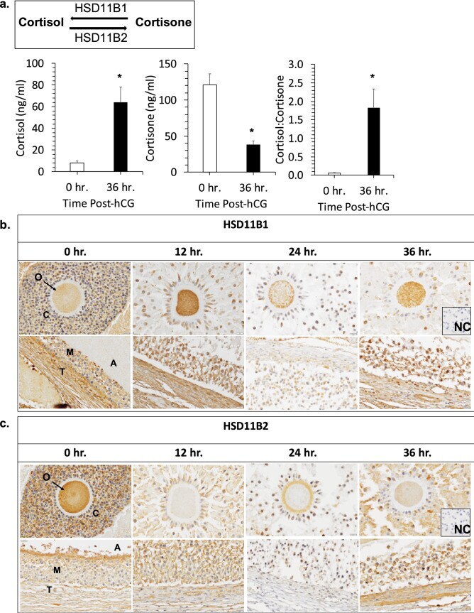
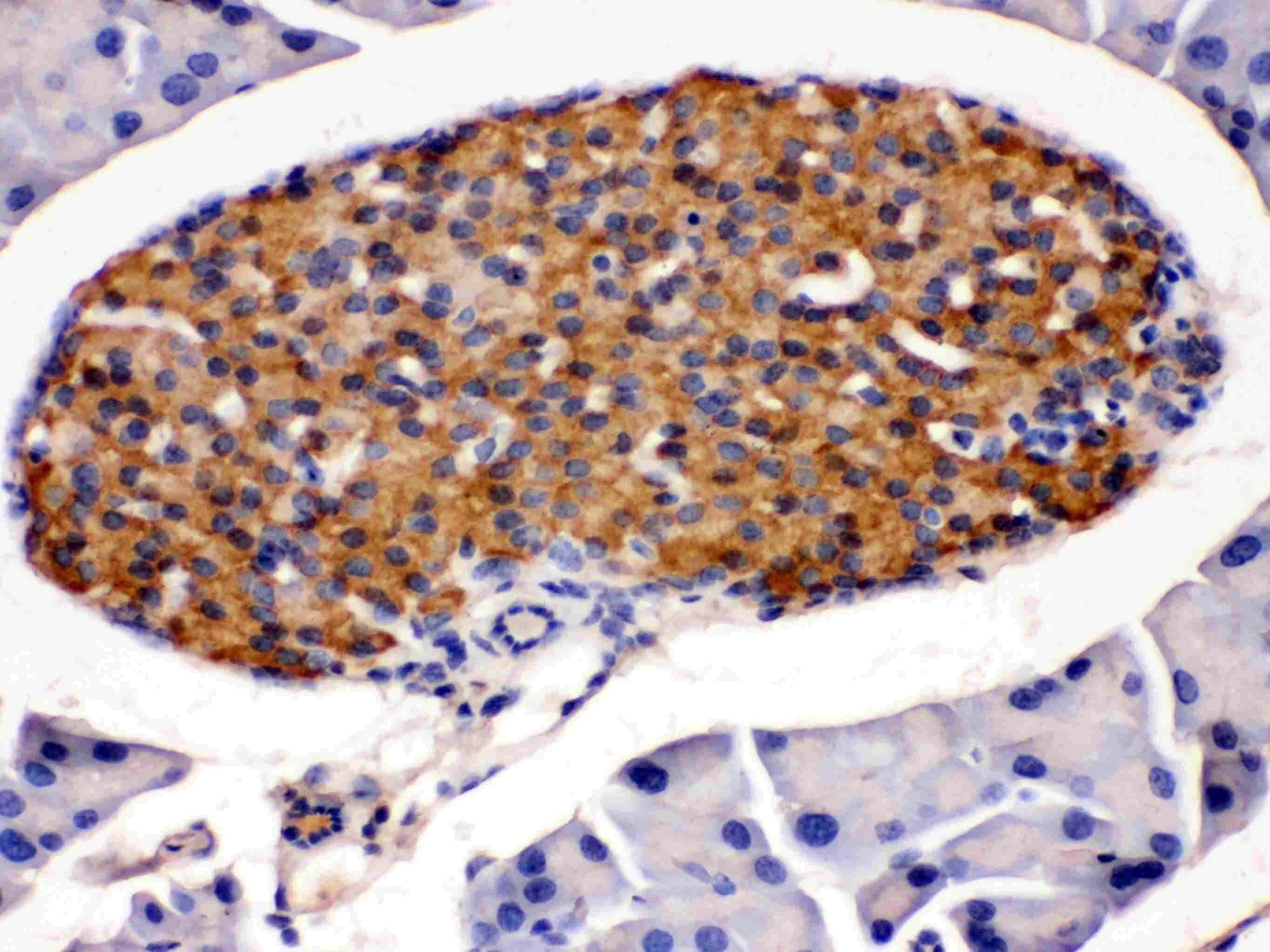
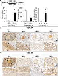
- Figure 4 Glucocorticoids and the enzymes that metabolize glucocorticoids are present in the rhesus macaque periovulatory follicle. ( a ) LC-MS/MS analysis of cortisol and cortisone concentrations in the FF obtained from rhesus macaque follicles between pre- (0 h) and 36 h post-hCG administration. A statistically significant increase in cortisol (* p = 0.0137) and decrease in cortisone (* p = 0.0110), with a corresponding increase in the cortisol to cortisone ratio (* p = 0.0256), was observed following hCG injection. ( b ) IHC of HSD11B1 and ( c ) HSD11B2 immunolocalization in the rhesus macaque periovulatory follicle pre- (0 h) as well as 12 h, 24 h and 36 h post-hCG administration. The images shown are representative of N = 4 ovaries obtained from separate animals undergoing a COv protocol at each of the times indicated. Note the overall increase in HSD11B1, as well as a concomitant decrease in HSD11B2 expression, in the ovarian follicle prior to hCG administration and with increasing time after the ovulatory stimulus. O = oocyte, C = cumulus cells, A = antrum, T = theca cells, M = mural granulosa cells and NC = negative control.