 Explore
 Validate
 Learn
Learn Western blot
 Immunocytochemistry
Antibody data
- Antibody Data
- Antigen structure
- References [6]
- Comments [0]
- Validations
- Immunocytochemistry [8]
- Chromatin Immunoprecipitation [4]
- Other assay [1]
Submit
Validation data
Reference
Comment
Report error
- Product number
- PA1-848 - Provider product page

- Provider
- Invitrogen Antibodies
- Product name
- p300 Polyclonal Antibody
- Antibody type
- Polyclonal
- Antigen
- Synthetic peptide
- Description
- PA1-848 detects p300 from human cells. PA1-848 has been successfully used in Western blot and ChIP procedures. By Western blot, this antibody detects an ~265 kDa protein representing p300 from HeLa cell lysate. The PA1-848 immunogen is a synthetic peptide corresponding to residues G(139) T S G P N Q G P T Q S T(151) C in the nuclear factor binding domain of human p300. The PA1-848 immunizing peptide (Cat. # PEP-053) is available for use in neutralization and control experiments.
- Reactivity
- Human
- Host
- Rabbit
- Isotype
- IgG
- Vial size
- 100 μg
- Concentration
- 1 mg/mL
- Storage
- -20°C, Avoid Freeze/Thaw Cycles
Submitted references Blocking elevated p38 MAPK restores efferocytosis and inflammatory resolution in the elderly.
MEF2D sustains activation of effector Foxp3+ Tregs during transplant survival and anticancer immunity.
DYRK1A interacts with histone acetyl transferase p300 and CBP and localizes to enhancers.
G Protein-coupled Receptor 40 (GPR40) and Peroxisome Proliferator-activated Receptor γ (PPARγ): AN INTEGRATED TWO-RECEPTOR SIGNALING PATHWAY.
Research resource: identification of novel coregulators specific for thyroid hormone receptor-β2.
Role of CBP/P300 in nuclear receptor signalling.
De Maeyer RPH, van de Merwe RC, Louie R, Bracken OV, Devine OP, Goldstein DR, Uddin M, Akbar AN, Gilroy DW
Nature immunology 2020 Jun;21(6):615-625
Nature immunology 2020 Jun;21(6):615-625
MEF2D sustains activation of effector Foxp3+ Tregs during transplant survival and anticancer immunity.
Di Giorgio E, Wang L, Xiong Y, Akimova T, Christensen LM, Han R, Samanta A, Trevisanut M, Bhatti TR, Beier UH, Hancock WW
The Journal of clinical investigation 2020 Dec 1;130(12):6242-6260
The Journal of clinical investigation 2020 Dec 1;130(12):6242-6260
DYRK1A interacts with histone acetyl transferase p300 and CBP and localizes to enhancers.
Li S, Xu C, Fu Y, Lei PJ, Yao Y, Yang W, Zhang Y, Washburn MP, Florens L, Jaiswal M, Wu M, Mohan M
Nucleic acids research 2018 Nov 30;46(21):11202-11213
Nucleic acids research 2018 Nov 30;46(21):11202-11213
G Protein-coupled Receptor 40 (GPR40) and Peroxisome Proliferator-activated Receptor γ (PPARγ): AN INTEGRATED TWO-RECEPTOR SIGNALING PATHWAY.
Wang S, Awad KS, Elinoff JM, Dougherty EJ, Ferreyra GA, Wang JY, Cai R, Sun J, Ptasinska A, Danner RL
The Journal of biological chemistry 2015 Aug 7;290(32):19544-57
The Journal of biological chemistry 2015 Aug 7;290(32):19544-57
Research resource: identification of novel coregulators specific for thyroid hormone receptor-β2.
Hahm JB, Privalsky ML
Molecular endocrinology (Baltimore, Md.) 2013 May;27(5):840-59
Molecular endocrinology (Baltimore, Md.) 2013 May;27(5):840-59
Role of CBP/P300 in nuclear receptor signalling.
Chakravarti D, LaMorte VJ, Nelson MC, Nakajima T, Schulman IG, Juguilon H, Montminy M, Evans RM
Nature 1996 Sep 5;383(6595):99-103
Nature 1996 Sep 5;383(6595):99-103
No comments: Submit comment
Supportive validation
- Submitted by
- Invitrogen Antibodies (provider)
- Main image
- Experimental details
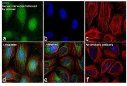
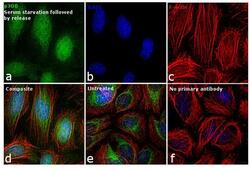
- Immunofluorescence analysis of p300 was performed using 70% confluent serum starved and released HeLa cells. The cells were fixed with 4% paraformaldehyde for 10 minutes, permeabilized with 0.1% Triton™ X-100 for 10 minutes, and blocked with 1% BSA for 1 hour at room temperature. The cells were labeled with p300 Rabbit polyclonal antibody (Product # PA1-848) at 5 µg/mL in 0.1% BSA, incubated overnight at 4 degree Celsius and then labeled with Goat anti-Rabbit IgG (H+L) Superclonal™ Secondary Antibody, Alexa Fluor® 488 conjugate (Product # A27034) at a dilution of 1:2000 for 45 minutes at room temperature (Panel a: green). Nuclei (Panel b: blue) were stained with SlowFade® Gold Antifade Mountant with DAPI (Product # S36938). F-actin (Panel c: red) was stained with Rhodamine Phalloidin (Product # R415, 1:300). Panel d represents the merged image showing nuclear localization. Panel e represents the untreated cells showing cytoplasmic localization of p300. Panel f shows control cells with no primary antibody to assess background. The images were captured at 60X magnification.
- Submitted by
- Invitrogen Antibodies (provider)
- Main image
- Experimental details
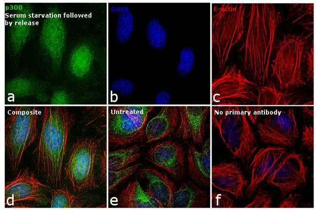
- Immunofluorescence analysis of p300 Polyclonal Antibody was performed using 70% confluent log phase HeLa cells. The cells were fixed with 4% paraformaldehyde for 10 minutes, permeabilized with 0.1% Triton™ X-100 for 15 minutes, and blocked with 2% BSA for 45 minutes at room temperature. The cells were labeled with p300 Polyclonal Antibody (Product # PA1-848) at 1:100 dilution in 0.1% BSA, incubated at 4 degree celsius overnight and then labeled with Donkey anti-Rabbit IgG (H+L) Highly Cross-Adsorbed Secondary Antibody, Alexa Fluor Plus 488 (Product # A32790), (1:2,000 dilution), for 45 minutes at room temperature (Panel a: Green). Nuclei (Panel b:Blue) were stained with ProLong™ Diamond Antifade Mountant with DAPI (Product # P36962). F-actin (Panel c: Red) was stained with Rhodamine Phalloidin (Product # R415, 1:300 dilution). Panel d represents the merged image showing nuclear localization. Panel e represents control cells with no primary antibody to assess background. The images were captured at 60X magnification.

- Submitted by
- Invitrogen Antibodies (provider)
- Main image

- Experimental details
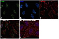
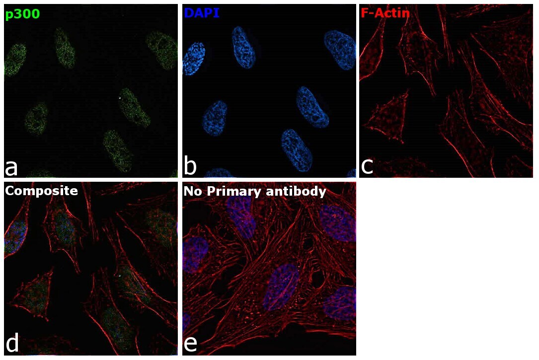
- Immunofluorescence analysis of p300 Polyclonal Antibody was performed using 70% confluent log phase HCT 116 cells. The cells were fixed with 4% paraformaldehyde for 10 minutes, permeabilized with 0.1% Triton™ X-100 for 15 minutes, and blocked with 2% BSA for 45 minutes at room temperature. The cells were labeled with p300 Polyclonal Antibody (Product # PA1-848) at 1:100 dilution in 0.1% BSA, incubated at 4 degree celsius overnight and then labeled with Donkey anti-Rabbit IgG (H+L) Highly Cross-Adsorbed Secondary Antibody, Alexa Fluor Plus 488 (Product # A32790), (1:2000 dilution), for 45 minutes at room temperature (Panel a: Green). Nuclei (Panel b:Blue) were stained with ProLong™ Diamond Antifade Mountant with DAPI (Product # P36962). F-actin (Panel c: Red) was stained with Rhodamine Phalloidin (Product # R415, 1:300 dilution). Panel d represents the merged image showing nuclear as well as cytoplasmic localization. Panel e represents control cells with no primary antibody to assess background. The images were captured at 60X magnification.(Ref: https://doi.org/10.1371/journal.pbio.3000732).
- Submitted by
- Invitrogen Antibodies (provider)
- Main image

- Experimental details
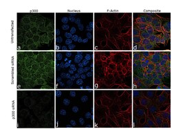
- Knockdown of p300 Polyclonal Antibody was achieved by transfecting HCT 116 cells with p300 specific siRNA (Silencer® select Product # S4696, S4697). Immunofluorescence analysis was performed on untransfected HCT 116 cells (panel a,d), transfected with non-specific scrambled siRNA (panels e,h) and transfected with p300 specific siRNA (panel i,l). Cells were fixed, permeabilized, and labelled with p300 Polyclonal Antibody (Product # PA1-848, 1:100 dilution) followed by Donkey anti-Rabbit IgG (H+L) Highly Cross-Adsorbed Secondary Antibody, Alexa Fluor Plus 488 (Product # A32790), (1:2000 dilution). Nuclei (blue) were stained using ProLong™ Diamond Antifade Mountant with DAPI (Product # P36962), and Rhodamine Phalloidin (Product # R415, 1:300 dilution) was used for cytoskeletal F-actin (Red) staining. reduction in signal was observed upon siRNA mediated knockdown (panel i,l) confirming specificity of the antibody to p300 (Green). The Images were captured at 60X magnification.

- Submitted by
- Invitrogen Antibodies (provider)
- Main image

- Experimental details
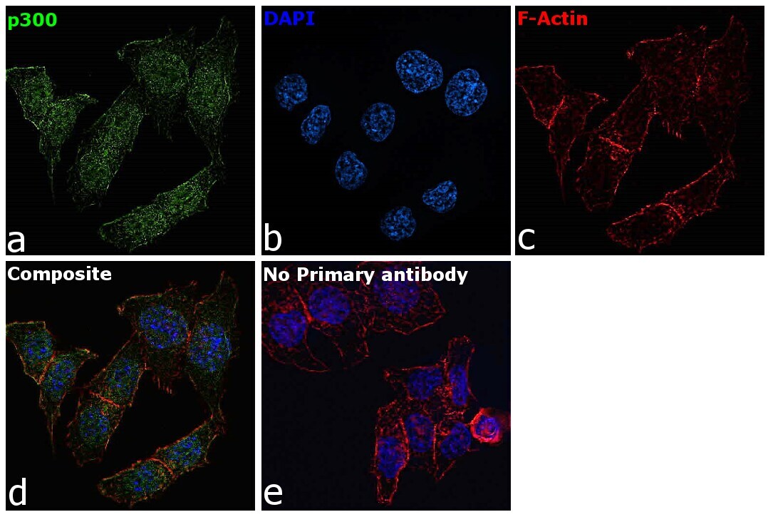
- Immunofluorescence analysis of p300 Polyclonal Antibody was performed using 70% confluent log phase HeLa cells. The cells were fixed with 4% paraformaldehyde for 10 minutes, permeabilized with 0.1% Triton™ X-100 for 15 minutes, and blocked with 2% BSA for 45 minutes at room temperature. The cells were labeled with p300 Polyclonal Antibody (Product # PA1-848) at 1:100 dilution in 0.1% BSA, incubated at 4 degree celsius overnight and then labeled with Donkey anti-Rabbit IgG (H+L) Highly Cross-Adsorbed Secondary Antibody, Alexa Fluor Plus 488 (Product # A32790), (1:2,000 dilution), for 45 minutes at room temperature (Panel a: Green). Nuclei (Panel b:Blue) were stained with ProLong™ Diamond Antifade Mountant with DAPI (Product # P36962). F-actin (Panel c: Red) was stained with Rhodamine Phalloidin (Product # R415, 1:300 dilution). Panel d represents the merged image showing nuclear localization. Panel e represents control cells with no primary antibody to assess background. The images were captured at 60X magnification.
- Submitted by
- Invitrogen Antibodies (provider)
- Main image

- Experimental details
- Immunofluorescence analysis of p300 Polyclonal Antibody was performed using 70% confluent log phase HCT 116 cells. The cells were fixed with 4% paraformaldehyde for 10 minutes, permeabilized with 0.1% Triton™ X-100 for 15 minutes, and blocked with 2% BSA for 45 minutes at room temperature. The cells were labeled with p300 Polyclonal Antibody (Product # PA1-848) at 1:100 dilution in 0.1% BSA, incubated at 4 degree celsius overnight and then labeled with Donkey anti-Rabbit IgG (H+L) Highly Cross-Adsorbed Secondary Antibody, Alexa Fluor Plus 488 (Product # A32790), (1:2000 dilution), for 45 minutes at room temperature (Panel a: Green). Nuclei (Panel b:Blue) were stained with ProLong™ Diamond Antifade Mountant with DAPI (Product # P36962). F-actin (Panel c: Red) was stained with Rhodamine Phalloidin (Product # R415, 1:300 dilution). Panel d represents the merged image showing nuclear as well as cytoplasmic localization. Panel e represents control cells with no primary antibody to assess background. The images were captured at 60X magnification.(Ref: https://doi.org/10.1371/journal.pbio.3000732).

- Submitted by
- Invitrogen Antibodies (provider)
- Main image
- Experimental details
- Knockdown of p300 Polyclonal Antibody was achieved by transfecting HCT 116 cells with p300 specific siRNA (Silencer® select Product # S4696, S4697). Immunofluorescence analysis was performed on untransfected HCT 116 cells (panel a,d), transfected with non-specific scrambled siRNA (panels e,h) and transfected with p300 specific siRNA (panel i,l). Cells were fixed, permeabilized, and labelled with p300 Polyclonal Antibody (Product # PA1-848, 1:100 dilution) followed by Donkey anti-Rabbit IgG (H+L) Highly Cross-Adsorbed Secondary Antibody, Alexa Fluor Plus 488 (Product # A32790), (1:2000 dilution). Nuclei (blue) were stained using ProLong™ Diamond Antifade Mountant with DAPI (Product # P36962), and Rhodamine Phalloidin (Product # R415, 1:300 dilution) was used for cytoskeletal F-actin (Red) staining. reduction in signal was observed upon siRNA mediated knockdown (panel i,l) confirming specificity of the antibody to p300 (Green). The Images were captured at 60X magnification.
- Submitted by
- Invitrogen Antibodies (provider)
- Main image
- Experimental details
- Immunofluorescence analysis of p300 was performed using 70% confluent serum starved and released HeLa cells. The cells were fixed with 4% paraformaldehyde for 10 minutes, permeabilized with 0.1% Triton™ X-100 for 10 minutes, and blocked with 1% BSA for 1 hour at room temperature. The cells were labeled with p300 Rabbit polyclonal antibody (Product # PA1-848) at 5 µg/mL in 0.1% BSA, incubated overnight at 4 degree Celsius and then labeled with Goat anti-Rabbit IgG (Heavy Chain) Superclonal™ Secondary Antibody, Alexa Fluor® 488 conjugate (Product # A27034) at a dilution of 1:2000 for 45 minutes at room temperature (Panel a: green). Nuclei (Panel b: blue) were stained with SlowFade® Gold Antifade Mountant with DAPI (Product # S36938). F-actin (Panel c: red) was stained with Rhodamine Phalloidin (Product # R415, 1:300). Panel d represents the merged image showing nuclear localization. Panel e represents the untreated cells showing cytoplasmic localization of p300. Panel f shows control cells with no primary antibody to assess background. The images were captured at 60X magnification.

Supportive validation
- Submitted by
- Invitrogen Antibodies (provider)
- Main image

- Experimental details
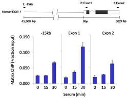
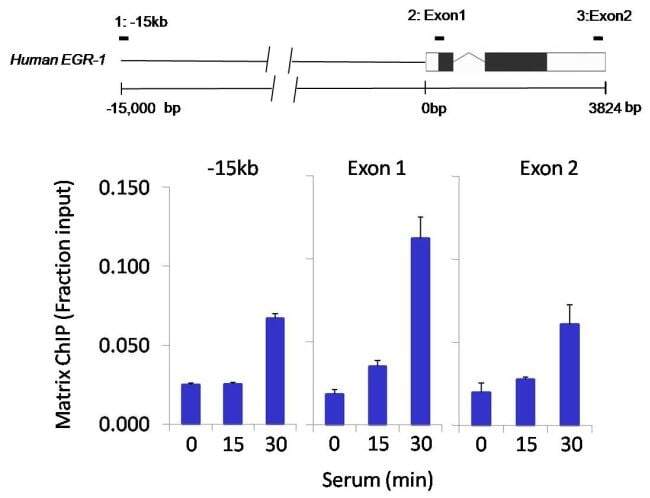
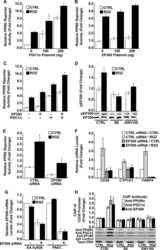
- Chromatin immunoprecipitation analysis of p300 was performed using cross-linked chromatin from 1x10^6 HCT116 colon carcinoma cells treated with serum for 0, 15, and 30 minutes. Immunoprecipitation was performed using a multiplex microplate Matrix ChIP assay (see reference for Matrix ChIP protocol: http://www.ncbi.nlm.nih.gov/pubmed/22098709) with 1.0 µL/100 µL well volume of a p300 polyclonal antibody (Product # PA1-848). Chromatin aliquots from ~1x10^5 cells were used per ChIP pull-down. Quantitative PCR data were done in quadruplicate using 1 µL of eluted DNA in 2 µL SYBR real-time PCR reactions containing primers to amplify -15kb upstream of the Egr1 gene or exon-1 or exon-2 of Egr1. PCR calibration curves were generated for each primer pair from a dilution series of sheared total genomic DNA. Quantitation of immunoprecipitated chromatin is presented as signal relative to the total amount of input chromatin. Results represent the mean +/- SEM for three experiments. A schematic representation of the Egr-1 locus is shown above the data where boxes represent exons (black boxes = translated regions, white boxes = untranslated regions), the zigzag line represents an intron, and the straight line represents upstream sequence. Regions amplified by Egr-1 primers are represented by black bars. Data courtesy of the Innovators Program.

- Submitted by
- Invitrogen Antibodies (provider)
- Main image

- Experimental details
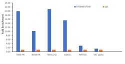
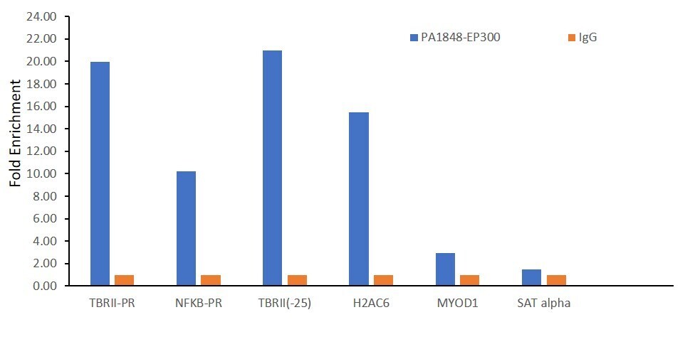
- Chromatin Immunoprecipitation (ChIP) was performed using p300 Polyclonal Antibody (Product # PA1-848, 5 µg) on sheared chromatin from a million HCT 116 cells using the MAGnify ChIP System (Product # 49-2024). Normal Rabbit IgG was used as a negative IP control. The purified DNA was analyzed by qPCR with PCR primer pairs over TGFβRII Promoter, TGFβRIIat -25K region, NFKB and H2AC genes (active), and MYOD1 and SAT Alpha (inactive). Antibody specificity was demonstrated by detection of enrichment of the target protein at specific gene loci. Data is presented as fold enrichment of the antibody signal versus the Rabbit Isotype using the comparative CT method.

- Submitted by
- Invitrogen Antibodies (provider)
- Main image
- Experimental details
- Chromatin immunoprecipitation analysis of p300 was performed using cross-linked chromatin from 1x10^6 HCT116 colon carcinoma cells treated with serum for 0, 15, and 30 minutes. Immunoprecipitation was performed using a multiplex microplate Matrix ChIP assay (see reference for Matrix ChIP protocol: http://www.ncbi.nlm.nih.gov/pubmed/22098709) with 1.0 µL/100 µL well volume of a p300 polyclonal antibody (Product # PA1-848). Chromatin aliquots from ~1x10^5 cells were used per ChIP pull-down. Quantitative PCR data were done in quadruplicate using 1 µL of eluted DNA in 2 µL SYBR real-time PCR reactions containing primers to amplify -15kb upstream of the Egr1 gene or exon-1 or exon-2 of Egr1. PCR calibration curves were generated for each primer pair from a dilution series of sheared total genomic DNA. Quantitation of immunoprecipitated chromatin is presented as signal relative to the total amount of input chromatin. Results represent the mean +/- SEM for three experiments. A schematic representation of the Egr-1 locus is shown above the data where boxes represent exons (black boxes = translated regions, white boxes = untranslated regions), the zigzag line represents an intron, and the straight line represents upstream sequence. Regions amplified by Egr-1 primers are represented by black bars. Data courtesy of the Innovators Program.
- Submitted by
- Invitrogen Antibodies (provider)
- Main image
- Experimental details
- Chromatin Immunoprecipitation (ChIP) was performed using p300 Polyclonal Antibody (Product # PA1-848, 5 µg) on sheared chromatin from a million HCT 116 cells using the MAGnify ChIP System (Product # 49-2024). Normal Rabbit IgG was used as a negative IP control. The purified DNA was analyzed by qPCR with PCR primer pairs over TGFβRII Promoter, TGFβRIIat -25K region, NFKB and H2AC genes (active), and MYOD1 and SAT Alpha (inactive). Antibody specificity was demonstrated by detection of enrichment of the target protein at specific gene loci. Data is presented as fold enrichment of the antibody signal versus the Rabbit Isotype using the comparative CT method.
Supportive validation
- Submitted by
- Invitrogen Antibodies (provider)
- Main image
- Experimental details
- NULL
