Explore
 Validate
 Learn
Learn Western blot
 Immunocytochemistry
Antibody data
- Antibody Data
- Antigen structure
- References [4]
- Comments [0]
- Validations
- Immunocytochemistry [3]
- Immunohistochemistry [2]
- Other assay [1]
Submit
Validation data
Reference
Comment
Report error
- Product number
- PA5-27399 - Provider product page

- Provider
- Invitrogen Antibodies
- Product name
- Adenosine Kinase Polyclonal Antibody
- Antibody type
- Polyclonal
- Antigen
- Recombinant full-length protein
- Description
- Recommended positive controls: A431, HepG2, HeLa, mouse liver, rat liver. Predicted reactivity: Mouse (89%), Rat (87%), Chicken (83%), Rhesus Monkey (99%), Bovine (93%). Store product as a concentrated solution. Centrifuge briefly prior to opening the vial.
- Reactivity
- Human, Mouse, Rat
- Host
- Rabbit
- Isotype
- IgG
- Vial size
- 100 μL
- Concentration
- 0.28 mg/mL
- Storage
- Store at 4°C short term. For long term storage, store at -20°C, avoiding freeze/thaw cycles.
Submitted references Role of Adenosine Kinase in Sphingosine-1-Phosphate Receptor 1-Induced Mechano-Hypersensitivities.
Adenosine Kinase Isoforms in the Developing Rat Hippocampus after LiCl/Pilocarpine Status Epilepticus.
Chronic Morphine-Induced Changes in Signaling at the A(3) Adenosine Receptor Contribute to Morphine-Induced Hyperalgesia, Tolerance, and Withdrawal.
Chemotherapy-induced pain is promoted by enhanced spinal adenosine kinase levels through astrocyte-dependent mechanisms.
Lauro F, Giancotti LA, Kolar G, Harada CM, Harmon TA, Garrett TJ, Salvemini D
Cellular and molecular neurobiology 2022 Nov;42(8):2909-2918
Cellular and molecular neurobiology 2022 Nov;42(8):2909-2918
Adenosine Kinase Isoforms in the Developing Rat Hippocampus after LiCl/Pilocarpine Status Epilepticus.
Fábera P, Uttl L, Kubová H, Tsenov G, Mareš P
International journal of molecular sciences 2022 Feb 24;23(5)
International journal of molecular sciences 2022 Feb 24;23(5)
Chronic Morphine-Induced Changes in Signaling at the A(3) Adenosine Receptor Contribute to Morphine-Induced Hyperalgesia, Tolerance, and Withdrawal.
Doyle TM, Largent-Milnes TM, Chen Z, Staikopoulos V, Esposito E, Dalgarno R, Fan C, Tosh DK, Cuzzocrea S, Jacobson KA, Trang T, Hutchinson MR, Bennett GJ, Vanderah TW, Salvemini D
The Journal of pharmacology and experimental therapeutics 2020 Aug;374(2):331-341
The Journal of pharmacology and experimental therapeutics 2020 Aug;374(2):331-341
Chemotherapy-induced pain is promoted by enhanced spinal adenosine kinase levels through astrocyte-dependent mechanisms.
Wahlman C, Doyle TM, Little JW, Luongo L, Janes K, Chen Z, Esposito E, Tosh DK, Cuzzocrea S, Jacobson KA, Salvemini D
Pain 2018 Jun;159(6):1025-1034
Pain 2018 Jun;159(6):1025-1034
No comments: Submit comment
Supportive validation
- Submitted by
- Invitrogen Antibodies (provider)
- Main image

- Experimental details
- Immunofluorescent analysis of Adenosine kinase in paraformaldehyde-fixed A431 cells using an Adenosine kinase polyclonal antibody (Product # PA5-27399) at a 1:200 dilution.
- Submitted by
- Invitrogen Antibodies (provider)
- Main image

- Experimental details
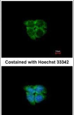
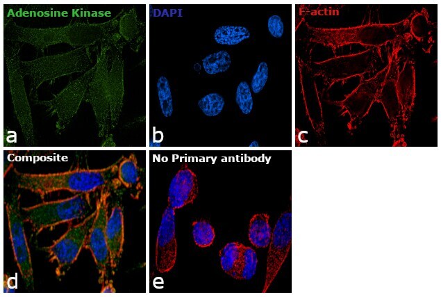
- Immunofluorescence analysis of Adenosine Kinase was performed using 70% confluent log phase PC-3 cells. The cells were fixed with 4% paraformaldehyde for 10 minutes, permeabilized with 0.1% Triton™ X-100 for 15 minutes, and blocked with 1% BSA for 1 hour at room temperature. The cells were labeled with Adenosine Kinase Polyclonal Antibody (Product # PA5-27399) at 5 µg/mL in 0.1% BSA, incubated at 4 degree Celsius overnight and then labeled with Goat anti-Rabbit IgG (H+L) Superclonal™ Secondary Antibody, Alexa Fluor® 488 conjugate (Product # A27034) at a dilution of 1:2000 for 45 minutes at room temperature (Panel a: green). Nuclei (Panel b: blue) were stained with ProLong™ Diamond Antifade Mountant with DAPI (Product # P36962). F-actin (Panel c: red) was stained with Rhodamine Phalloidin (Product # R415, 1:300). Panel d represents the merged image showing cytoplasmic localization. Panel e represents control cells with no primary antibody to assess background. The images were captured at 60X magnification.

- Submitted by
- Invitrogen Antibodies (provider)
- Main image

- Experimental details
- Immunofluorescence analysis of Adenosine Kinase was performed using 70% confluent log phase PC-3 cells. The cells were fixed with 4% paraformaldehyde for 10 minutes, permeabilized with 0.1% Triton™ X-100 for 15 minutes, and blocked with 1% BSA for 1 hour at room temperature. The cells were labeled with Adenosine Kinase Polyclonal Antibody (Product # PA5-27399) at 5 µg/mL in 0.1% BSA, incubated at 4 degree Celsius overnight and then labeled with Goat anti-Rabbit IgG (Heavy Chain) Superclonal™ Secondary Antibody, Alexa Fluor® 488 conjugate (Product # A27034) at a dilution of 1:2000 for 45 minutes at room temperature (Panel a: green). Nuclei (Panel b: blue) were stained with ProLong™ Diamond Antifade Mountant with DAPI (Product # P36962). F-actin (Panel c: red) was stained with Rhodamine Phalloidin (Product # R415, 1:300). Panel d represents the merged image showing cytoplasmic localization. Panel e represents control cells with no primary antibody to assess background. The images were captured at 60X magnification.
Supportive validation
- Submitted by
- Invitrogen Antibodies (provider)
- Main image
- Experimental details
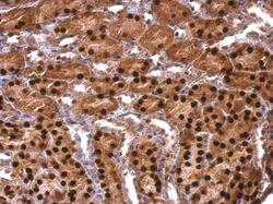
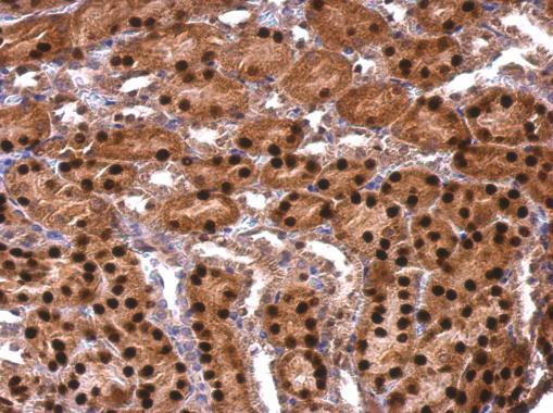
- Adenosine Kinase Polyclonal Antibody detects Adenosine kinase protein at nucleus on mouse kidney by immunohistochemical analysis. Sample: Paraffin-embedded mouse kidney. Adenosine Kinase Polyclonal Antibody (Product # PA5-27399) dilution: 1:500. Antigen Retrieval: EDTA based buffer, pH 8.0, 15 min.
- Submitted by
- Invitrogen Antibodies (provider)
- Main image
- Experimental details
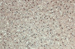
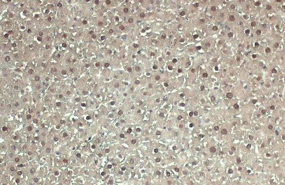
- Adenosine Kinase Polyclonal Antibody detects ADK protein at cytoplasm and nucleus by immunohistochemical analysis. Sample: Paraffin-embedded mouse liver. ADK stained by Adenosine Kinase Polyclonal Antibody (Product # PA5-27399) diluted at 1:500. Antigen Retrieval: Citrate buffer, pH 6.0, 15 min.
Supportive validation
- Submitted by
- Invitrogen Antibodies (provider)
- Main image
- Experimental details
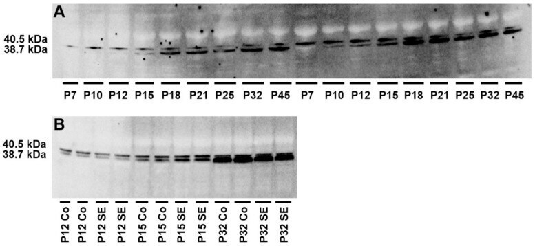
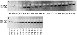
- Western blotting of hippocampal homogenates showing the density of ADK isoforms-ADK-S (38.7 kDa) and ADK-L (40.5 kDa) during brain development in control rats ( A ) and corresponding age-matched LiCl/pilocarpine SE rats ( B ). ( A ) Abscissae from left to right: 7, 10, 12, 15, 18, 21, 25, 32, and 45-day-old rats in identical replicates. ( B ) Abscissae from left to right: 12-, 15-, and 32-day-old rats in identical replicates.