 Explore
 Validate
 Learn
Learn Western blot
Antibody data
- Antibody Data
- Antigen structure
- References [1]
- Comments [0]
- Validations
- Western blot [1]
- Immunocytochemistry [5]
- Immunohistochemistry [2]
- Other assay [1]
Submit
Validation data
Reference
Comment
Report error
- Product number
- PA5-20813 - Provider product page

- Provider
- Invitrogen Antibodies
- Product name
- AGTR2 Polyclonal Antibody
- Antibody type
- Polyclonal
- Antigen
- Synthetic peptide
- Description
- A suggested positive control is mouse liver tissue lysate. PA5-20813 can be used with blocking peptide PEP-0927.
- Reactivity
- Human, Mouse, Rat
- Host
- Rabbit
- Isotype
- IgG
- Vial size
- 100 μg
- Concentration
- 1 mg/mL
- Storage
- Maintain refrigerated at 2-8°C for up to 3 months. For long term storage store at -20°C
Submitted references Relaxin Inhibits the Cardiac Myofibroblast NLRP3 Inflammasome as Part of Its Anti-Fibrotic Actions via the Angiotensin Type 2 and ATP (P2X7) Receptors.
Tapia Cáceres F, Gaspari TA, Hossain MA, Samuel CS
International journal of molecular sciences 2022 Jun 25;23(13)
International journal of molecular sciences 2022 Jun 25;23(13)
No comments: Submit comment
Supportive validation
- Submitted by
- Invitrogen Antibodies (provider)
- Main image
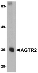
- Experimental details
- Western Blot analysis of AGTR2 in mouse liver tissue lysate with AGTR2 Polyclonal Antibody (Product # PA5-20813) at 0.5 µg/mL.

Supportive validation
- Submitted by
- Invitrogen Antibodies (provider)
- Main image
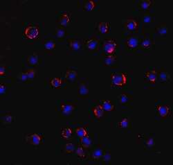
- Experimental details
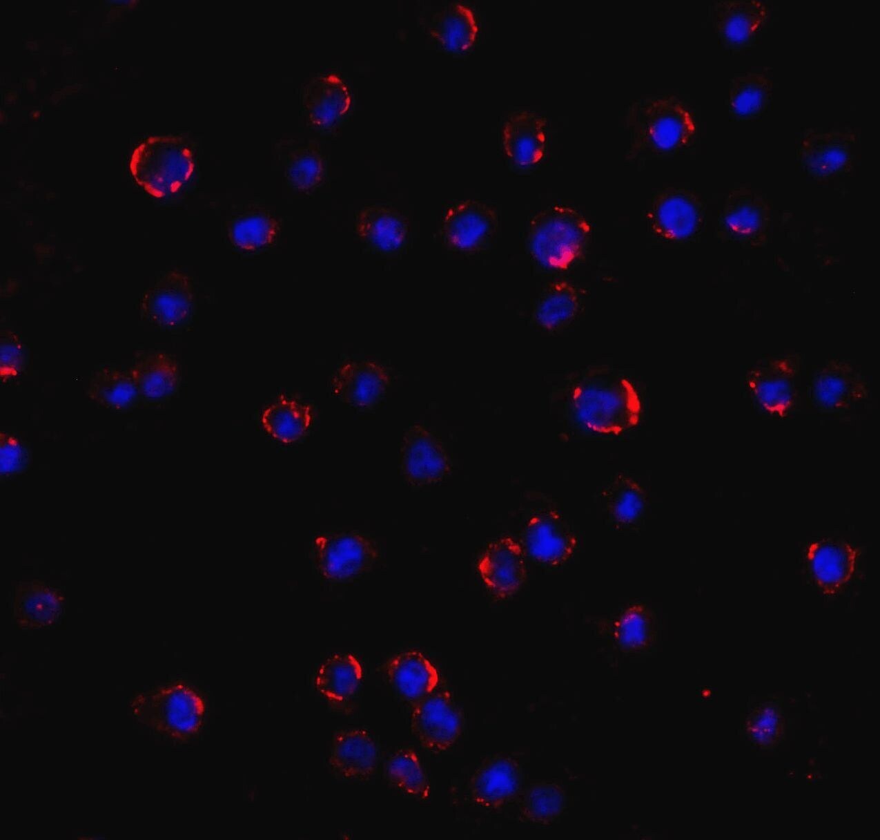
- Immunofluorescence of AGTR2 in Jurkat cell with AGTR2 Polyclonal Antibody (Product # PA5-20813) at 5 µg/mL.
- Submitted by
- Invitrogen Antibodies (provider)
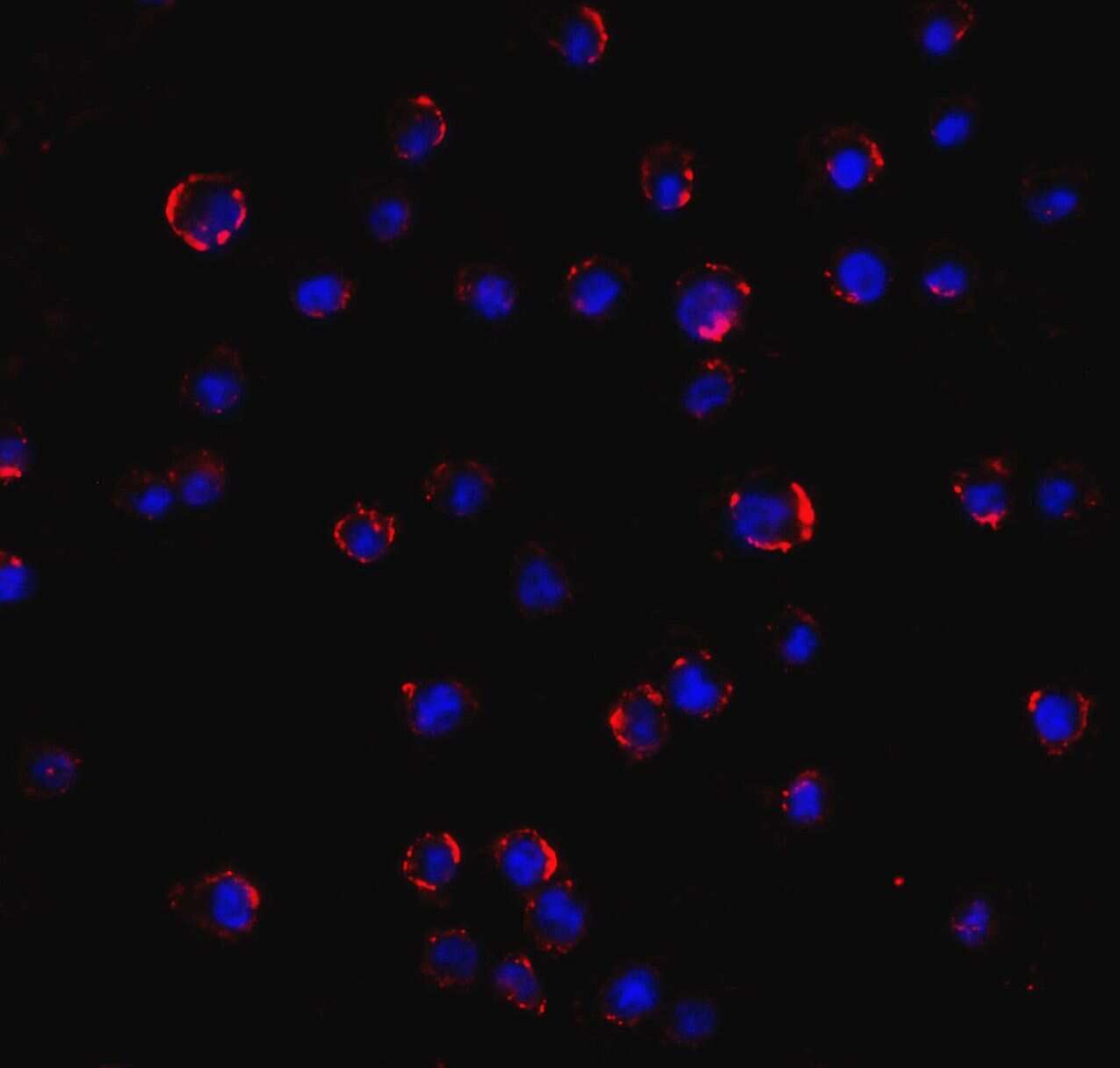
- Main image

- Experimental details
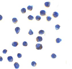
- Immunocytochemistry of AGTR2 in Jurkat cell with AGTR2 Polyclonal Antibody (Product # PA5-20813) at 1 µg/mL.

- Submitted by
- Invitrogen Antibodies (provider)
- Main image
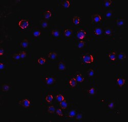
- Experimental details
- Immunofluorescence of AGTR2 in Jurkat cell with AGTR2 Polyclonal Antibody (Product # PA5-20813) at 5 µg/mL.

- Submitted by
- Invitrogen Antibodies (provider)
- Main image

- Experimental details
- Immunofluorescence of AGTR2 in Jurkat cell with AGTR2 Polyclonal Antibody (Product # PA5-20813) at 5 µg/mL.
- Submitted by
- Invitrogen Antibodies (provider)
- Main image
- Experimental details
- Immunocytochemistry of AGTR2 in Jurkat cell with AGTR2 Polyclonal Antibody (Product # PA5-20813) at 1 µg/mL.

Supportive validation
- Submitted by
- Invitrogen Antibodies (provider)
- Main image
- Experimental details
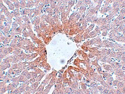
- Immunohistochemistry of AGTR2 in rat liver tissue with AGTR2 Polyclonal Antibody (Product # PA5-20813) at 5 µg/mL.
- Submitted by
- Invitrogen Antibodies (provider)
- Main image

- Experimental details
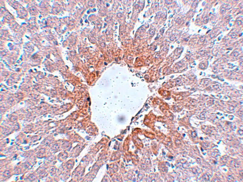
- Immunohistochemistry of AGTR2 in rat liver tissue with AGTR2 Polyclonal Antibody (Product # PA5-20813) at 5 µg/mL.

Supportive validation
- Submitted by
- Invitrogen Antibodies (provider)
- Main image
- Experimental details
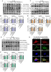
- The effects of RLX +- the RXFP1 inhibitor (RXFP1-I), AT 2 R inhibitor (PD) or P2X7R inhibitor (A4) on the T+L+Bz-stimulated increase in TLR-4, NLRP3, pro-caspase-1, pro-IL-1beta and pro-IL-18 levels from HCMFs. ( A , C , E ) Representative Western blots of TLR-4, NLRP3, pro-caspase-1, pro-IL-beta and pro-IL-8 expression from HCMFs treated with TGF-beta1 (T), T+L+Bz, T+L+Bz+RXFP1-I, T+L+Bz+RLX or T+L+Bz+RLX+RXFP1-I ( A ); T, T+L+Bz, T+L+Bz+PD, T+L+Bz+RLX or T+L+Bz+RLX+PD ( C ); or T, T+L+Bz, T+L+Bz+A4, T+L+Bz+RLX or T+L+Bz+RLX+A4 ( E ), after 8 h. ( B , D , F ) Also shown are the mean +- SEM of each end point measured from each treatment group studied, corrected for GAPDH (house-keeping protein) loading, and expressed relative to the value in the T-stimulated cell group, which was expressed as 1 in each case, from n =6 separate experiments conducted in duplicate. The individual data sets for each group are represented by the white circles within each set of bar graphs. ( G ) Representative immunofluorescence staining of RXFP1, AT 2 R and P2X7R, and colocalization of RXFP1 and P2X7R or AT 2 R and P2X7R in HCMFs. Scale bar = 20 mum. Nuclear staining was performed using DAPI, whilst merged images demonstrated that all three receptors were expressed by HCMFs. * p < 0.05, ** p < 0.01 compared to the T alone group; # p < 0.05 compared to the T+L+Bz group; P p < 0.05 compared to the T+L+Bz+RLX group.
