Explore
 Validate
 Learn
Learn Western blot
 Immunohistochemistry
Antibody data
- Antibody Data
- Antigen structure
- References [1]
- Comments [0]
- Validations
- Immunohistochemistry [1]
Submit
Validation data
Reference
Comment
Report error
- Product number
- A02423-2 - Provider product page

- Provider
- Boster Biological Technology
- Product name
- Anti-Bag1 Antibody Picoband™
- Antibody type
- Polyclonal
- Description
- Polyclonal antibody for Bag-1/BAG1 detection. Host: Rabbit.Size: 100μg/vial. Tested applications: IHC-P. Reactive species: Human. Bag-1/BAG1 information: Molecular Weight: 38779 MW; Subcellular Localization: Isoform 1: Nucleus. Cytoplasm. Isoform 1 localizes predominantly to the nucleus; Tissue Specificity: Isoform 4 is the most abundantly expressed isoform. It is ubiquitously expressed throughout most tissues, except the liver, colon, breast and uterine myometrium. Isoform 1 is expressed in the ovary and testis. Isoform 4 is expressed in several types of tumor cell lines, and at consistently high levels in leukemia and lymphoma cell lines. Isoform 1 is expressed in the prostate, breast and leukemia cell lines. Isoform 3 is the least abundant isoform in tumor cell lines (at protein level).
- Reactivity
- Human, Mouse, Rat
- Host
- Rabbit
- Vial size
- 100μg/vial
- Concentration
- Add 0.2ml of distilled water will yield a concentration of 500ug/ml.
- Storage
- At -20°C for one year. After reconstitution, at 4°C for one month. It can also be aliquoted and stored frozen at -20°C for a longer time. Avoid repeated freezing and thawing.
- Handling
- Add 0.2ml of distilled water will yield a concentration of 500ug/ml.
Submitted references Clinicopathologic significance of BAG1 and TIMP3 expression in colon carcinoma.
Bai YX, Yi JL, Li JF, Sui H
World journal of gastroenterology 2007 Jul 28;13(28):3883-5
World journal of gastroenterology 2007 Jul 28;13(28):3883-5
No comments: Submit comment
Supportive validation
- Submitted by
- Boster Biological Technology (provider)
- Main image
- Experimental details
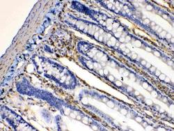
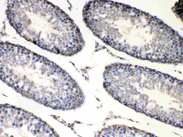
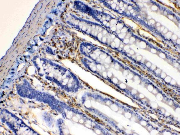
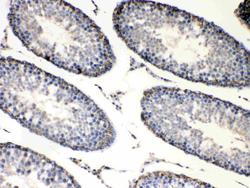
- IHC analysis of Bag1 using anti-Bag1 antibody (A02423-2). Bag1 was detected in paraffin-embedded section of rat testis tissues. Heat mediated antigen retrieval was performed in citrate buffer (pH6, epitope retrieval solution) for 20 mins. The tissue section was blocked with 10% goat serum. The tissue section was then incubated with 1μg/ml rabbit anti- Bag1 Antibody (A02423-2) overnight at 4°C. Biotinylated goat anti-rabbit IgG was used as secondary antibody and incubated for 30 minutes at 37°C. The tissue section was developed using Strepavidin-Biotin-Complex (SABC)(Catalog # SA1022) with DAB as the chromogen.
- Additional image