 Explore
 Validate
 Learn
Learn Western blot
 Immunocytochemistry
Antibody data
- Antibody Data
- Antigen structure
- References [1]
- Comments [0]
- Validations
- Immunocytochemistry [1]
Submit
Validation data
Reference
Comment
Report error
- Product number
- HPA051019 - Provider product page

- Provider
- Atlas Antibodies
- Proper citation
- Atlas Antibodies Cat#HPA051019, RRID:AB_2681313
- Product name
- Anti-ARFGAP1
- Antibody type
- Polyclonal
- Description
- Polyclonal Antibody against Human ARFGAP1, Gene description: ADP-ribosylation factor GTPase activating protein 1, Alternative Gene Names: ARF1GAP, bA261N11.3, FLJ10767, Validated applications: WB, IHC, ICC, Uniprot ID: Q8N6T3, Storage: Store at +4°C for short term storage. Long time storage is recommended at -20°C.
- Reactivity
- Human
- Host
- Rabbit
- Conjugate
- Unconjugated
- Isotype
- IgG
- Vial size
- 100 µl
- Concentration
- 0.2 mg/ml
- Storage
- Store at +4°C for short term storage. Long time storage is recommended at -20°C.
- Handling
- The antibody solution should be gently mixed before use.
Submitted references An ARF GTPase module promoting invasion and metastasis through regulating phosphoinositide metabolism
Nacke M, Sandilands E, Nikolatou K, Román-Fernández Á, Mason S, Patel R, Lilla S, Yelland T, Galbraith L, Freckmann E, McGarry L, Morton J, Shanks E, Leung H, Markert E, Ismail S, Zanivan S, Blyth K, Bryant D
Nature Communications 2021;12(1)
Nature Communications 2021;12(1)
No comments: Submit comment
Supportive validation
- Submitted by
- Atlas Antibodies (provider)
- Main image
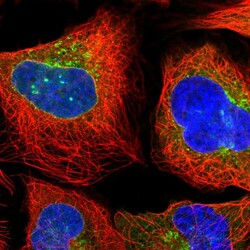
- Experimental details
- Immunofluorescent staining of human cell line U-2 OS shows localization to nuclear membrane, cytosol, the Golgi apparatus & vesicles.
- Sample type
- Human
