 Explore
 Validate
 Learn
Learn Western blot
 Immunocytochemistry
Antibody data
- Antibody Data
- Antigen structure
- References [1]
- Comments [0]
- Validations
- Immunocytochemistry [1]
Submit
Validation data
Reference
Comment
Report error
- Product number
- MA1-830 - Provider product page

- Provider
- Invitrogen Antibodies
- Product name
- PLA2G5 Monoclonal Antibody (3G1)
- Antibody type
- Monoclonal
- Antigen
- Other
- Description
- MA1-830 contains 100 µg of in vitro produced, protein A purified antibody (1 mg/mL) in PBS containing 1 mg/mL BSA and 0.05% sodium azide. MA1-830 detects Group V Secretory Phospholipase A2 from human and mouse samples. MA1-830 has been successfully used in Western blot procedures. By Western blot, MA1-830 detects a ~15 kDa band representing Group V Secretory Phospholipase A2 from mouse macrophages. The MA1-830 immunogen is full length W79A human group V phospholipase A2.
- Reactivity
- Human, Mouse
- Host
- Mouse
- Isotype
- IgG
- Antibody clone number
- 3G1
- Vial size
- 100 μg
- Concentration
- 1 mg/mL
- Storage
- -20°C, Avoid Freeze/Thaw Cycles
Submitted references Characterization of monoclonal antibodies specific for 14-kDa human group V secretory phospholipase A2 (hVPLA2).
Muñoz NM, Kim KP, Han SK, Boetticher E, Sperling AI, Sano H, Zhu X, Cho W, Leff AR
Hybridoma 2000 Apr;19(2):171-6
Hybridoma 2000 Apr;19(2):171-6
No comments: Submit comment
Supportive validation
- Submitted by
- Invitrogen Antibodies (provider)
- Main image
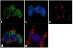
- Experimental details
- Immunofluorescence analysis of Group V secretory phospholipase A2 (PLAG5) was performed using 90% confluent log phase NCCIT cells. The cells were fixed with 4% paraformaldehyde for 10 minutes, permeabilized with 0.1% Triton™ X-100 for 10 minutes, and blocked with 1% BSA for 1 hour at room temperature. The cells were labeled with PLA2G5 (3G1) Mouse Monoclonal Antibody (Product # MA1-830) at 2µg/mL in 0.1% BSA and incubated for 3 hours at room temperature and then labeled with Goat anti-Mouse IgG (H+L) Superclonal™ Secondary Antibody, Alexa Fluor® 488 conjugate (Product # A28175) at a dilution of 1:2000 for 45 minutes at room temperature (Panel a: green). Nuclei (Panel b: blue) were stained with SlowFade® Gold Antifade Mountant with DAPI (Product # S36938). F-actin (Panel c: red) was stained with Alexa Fluor® 555 Rhodamine Phalloidin (Product # R415, 1:300). Panel d represents the merged image showing cytoplasmic localization. Panel e shows the no primary antibody control. The images were captured at 60X magnification.
