Explore
 Validate
 Learn
Learn Western blot
 Immunocytochemistry
Antibody data
- Antibody Data
- Antigen structure
- References [4]
- Comments [0]
- Validations
- Immunocytochemistry [1]
Submit
Validation data
Reference
Comment
Report error
- Product number
- HPA014059 - Provider product page

- Provider
- Atlas Antibodies
- Proper citation
- Atlas Antibodies Cat#HPA014059, RRID:AB_1858029
- Product name
- Anti-SEC62
- Antibody type
- Polyclonal
- Description
- Polyclonal Antibody against Human SEC62, Gene description: SEC62 homolog (S. cerevisiae), Alternative Gene Names: Dtrp1, HTP1, TLOC1, Validated applications: WB, IHC, ICC, Uniprot ID: Q99442, Storage: Store at +4°C for short term storage. Long time storage is recommended at -20°C.
- Reactivity
- Human
- Host
- Rabbit
- Conjugate
- Unconjugated
- Isotype
- IgG
- Vial size
- 100 µl
- Concentration
- 0.1 mg/ml
- Storage
- Store at +4°C for short term storage. Long time storage is recommended at -20°C.
- Handling
- The antibody solution should be gently mixed before use.
Submitted references ER entry pathway and glycosylation of GPI-anchored proteins are determined by N-terminal signal sequence and C-terminal GPI-attachment sequence
Rab32 uses its effector reticulon 3L to trigger autophagic degradation of mitochondria-associated membrane (MAM) proteins
Mammalian SRP receptor switches the Sec61 translocase from Sec62 to SRP-dependent translocation
Systematic Interrogation of 3q26 Identifies TLOC1 and SKIL as Cancer Drivers
Hirata T, Yang J, Tomida S, Tokoro Y, Kinoshita T, Fujita M, Kizuka Y
Journal of Biological Chemistry 2022;298(10):102444
Journal of Biological Chemistry 2022;298(10):102444
Rab32 uses its effector reticulon 3L to trigger autophagic degradation of mitochondria-associated membrane (MAM) proteins
Herrera-Cruz M, Yap M, Tahbaz N, Phillips K, Thomas L, Thomas G, Simmen T
Biology Direct 2021;16(1)
Biology Direct 2021;16(1)
Mammalian SRP receptor switches the Sec61 translocase from Sec62 to SRP-dependent translocation
Jadhav B, McKenna M, Johnson N, High S, Sinning I, Pool M
Nature Communications 2015;6(1)
Nature Communications 2015;6(1)
Systematic Interrogation of 3q26 Identifies TLOC1 and SKIL as Cancer Drivers
Hagerstrand D, Tong A, Schumacher S, Ilic N, Shen R, Cheung H, Vazquez F, Shrestha Y, Kim S, Giacomelli A, Rosenbluh J, Schinzel A, Spardy N, Barbie D, Mermel C, Weir B, Garraway L, Tamayo P, Mesirov J, Beroukhim R, Hahn W
Cancer Discovery 2013;3(9):1044-1057
Cancer Discovery 2013;3(9):1044-1057
No comments: Submit comment
Supportive validation
- Submitted by
- Atlas Antibodies (provider)
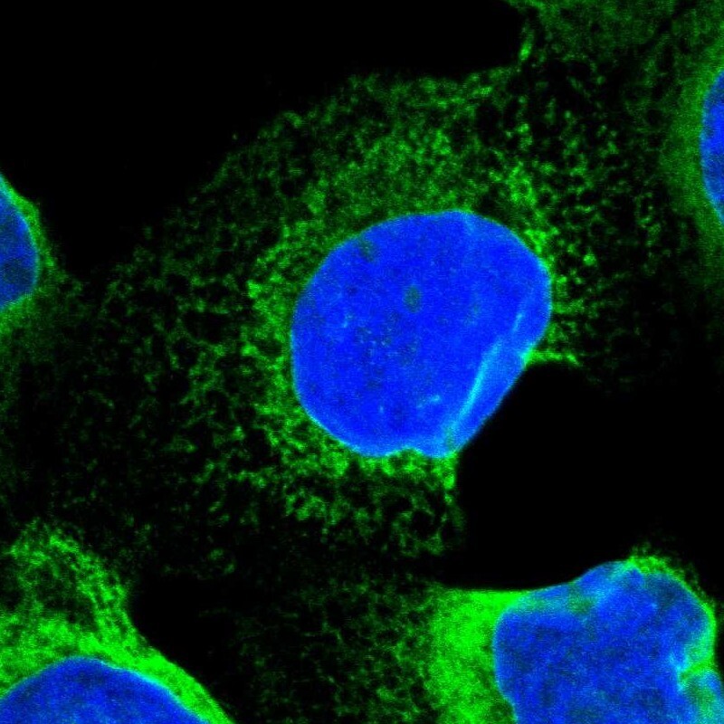
- Main image
- Experimental details
- Immunofluorescent staining of human cell line A-431 shows positivity in endoplasmic reticulum.
- Sample type
- Human