Explore
 Validate
 Learn
Learn Western blot
Antibody data
- Antibody Data
- Antigen structure
- References [0]
- Comments [0]
- Validations
- Western blot [1]
- Immunoprecipitation [1]
- Immunohistochemistry [1]
Submit
Validation data
Reference
Comment
Report error
- Product number
- MA5-55962 - Provider product page

- Provider
- Invitrogen Antibodies
- Product name
- CDA Monoclonal Antibody (K1E006_15H12)
- Antibody type
- Monoclonal
- Antigen
- Recombinant full-length protein
- Description
- Sequence of this protein is as follows: MAQKRPACTL KPECVQQLLV CSQEAKKSAY CPYSHFPVGA ALLTQEGRIF KGCNIENACY PLGICAERTA IQKAVSEGYK DFRAIAIASD MQDDFISPCG ACRQVMREFG TNWPVYMTKP DGTYIVMTVQ ELLPSSFGPE DLQKTQ
- Reactivity
- Human
- Host
- Mouse
- Isotype
- IgG
- Antibody clone number
- K1E006_15H12
- Vial size
- 50 μg
- Concentration
- 1 mg/mL
- Storage
- Store at 4°C short term. For long term storage, store at -20°C, avoiding freeze/thaw cycles.
No comments: Submit comment
Supportive validation
- Submitted by
- Invitrogen Antibodies (provider)
- Main image
- Experimental details
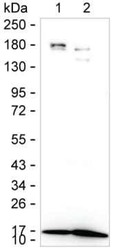
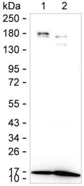
- Western blot analysis of CDA in 15 µg of SK-OV-3 lysate. Sample was run on 6-18% SDS-PAGE under reducing conditions, blotted onto nitrocellulose membrane, and peroxidase conjugated goat anti-mouse IgG was used as the secondary antibody. CDA band was visualized using ECL Substrate. Incubation with primary CDA monoclonal antibody (Product # MA5-55962) at a dilution of 1 µg/mL was used.
Supportive validation
- Submitted by
- Invitrogen Antibodies (provider)
- Main image
- Experimental details
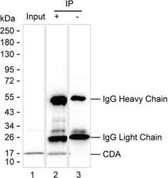
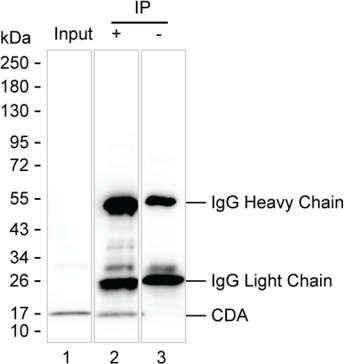
- Immunoprecipitation of CDA in 200 µg of SK-OV-3 lysate. Samples are as follows: Lane 1: SK-OV-3 lysate, Lane 2: CDK4 immunoprecipitated from SK-OV-3 lysate, Lane3: The same as Lane 2 but KT82 was used as IgG isotype control antibody. After absorption with Protein G beads, the mixture was run on 6-18% SDS-PAGE, blotted onto nitrocellulose membrane, and peroxidase conjugated goat anti-mouse IgG was used as the secondary antibody. The isotype control antibody was KT82. Incubation of samples with CDA monoclonal antibody (Product # MA5-55962) at a dilution of 2.5 µg was used.
Supportive validation
- Submitted by
- Invitrogen Antibodies (provider)
- Main image

- Experimental details
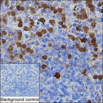
- Immunohistochemistry analysis of CDA in paraffin-embedded spleen tissue. Sample was incubated with CDA monoclonal antibody (Product # MA5-55962) at a dilution of 1 µg/mL (RT, 1 hour). Antigen was retrieved through addition of boiling Tris/EDTA buffer pH 9 in a pressure cooker for 3 min. Endogenous peroxidase activity was quenched by incubating the sections with 3% H2O2 for 30 min at room temperature. Poly-peroxidase conjugated goat anti-mouse IgG was used as the secondary antibody. Diaminobenzidine was used as the chromogen. The section was counterstained with hematoxylin. A tissue section incubated with phosphate-buffered saline followed by incubation with the secondary antibody was used as the background control.