Explore
 Validate
 Learn
Learn Western blot
 Immunoprecipitation
Antibody data
- Antibody Data
- Antigen structure
- References [1]
- Comments [0]
- Validations
- Western blot [2]
- Immunohistochemistry [1]
- Other assay [4]
Submit
Validation data
Reference
Comment
Report error
- Product number
- PA5-80305 - Provider product page

- Provider
- Invitrogen Antibodies
- Product name
- ACSS3 Polyclonal Antibody
- Antibody type
- Polyclonal
- Antigen
- Synthetic peptide
- Description
- For reconstitution, we recommend adding 100 µL distilled water to a final antibody concentration of about 1 mg/mL. To use this carrier-free antibody for conjugation experiments, we strongly recommend performing another round of desalting. (Zeba Spin Desalting Columns, 7KMWCO, 0.5 mL, Product # 89882)
- Reactivity
- Human
- Host
- Rabbit
- Isotype
- IgG
- Vial size
- 100 µL
- Concentration
- 1 mg/mL
- Storage
- Maintain refrigerated at 2-8°C for up to 1 month. For long term storage store at -20°C
Submitted references Acetyl-CoA synthetase 3 promotes bladder cancer cell growth under metabolic stress.
Zhang J, Duan H, Feng Z, Han X, Gu C
Oncogenesis 2020 May 12;9(5):46
Oncogenesis 2020 May 12;9(5):46
No comments: Submit comment
Supportive validation
- Submitted by
- Invitrogen Antibodies (provider)
- Main image

- Experimental details
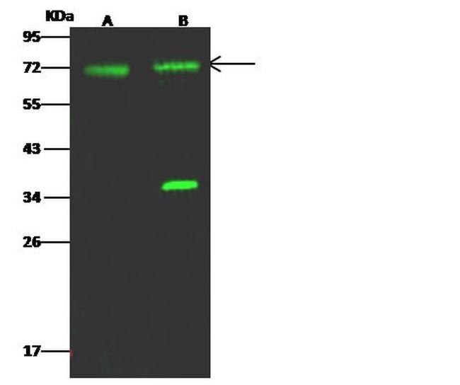
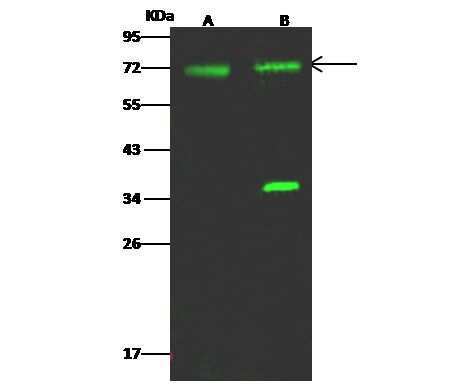
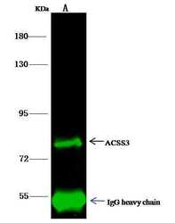
- Western blot analysis of ACSS3 in Lane A: HepG2 Whole Cell lysate (30 µg), Lane B: MCF7 Whole Cell lysate (30 µg). Samples were probed using a ACSS3 Polyclonal Antibody (Product # PA5-80305) at a 1:500 dilution, followed by a Goat Anti-Rabbit IgG (H+L), Dylight 800 Secondary Antibody at a 1:10000 dilution. Western blot was performed under reducing conditions. Predicted band size:75 kDa. Observed band size:72 kDa.
- Submitted by
- Invitrogen Antibodies (provider)
- Main image
- Experimental details
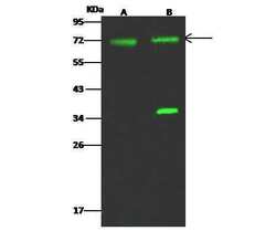
- Western Blot using ACSS3 Polyclonal Antibody (Product # PA5-80305) at 1:500 dilution. Lane A: HepG2 Whole Cell Lysate, Lane B: MCF7 Whole Cell Lysate. Lysates/proteins at 30 μg per lane. Secondary antibody: Goat Anti- Rabbit IgG H&L (DyLight™ 800) at 1:10,000 dilution. Developed using the Odyssey technique. Performed under reducing conditions. Predicted band size: 75 kDa. Observed band size: 72 kDa. (We are unsure of the identity of these extra bands).
Supportive validation
- Submitted by
- Invitrogen Antibodies (provider)
- Main image
- Experimental details
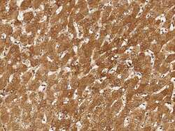
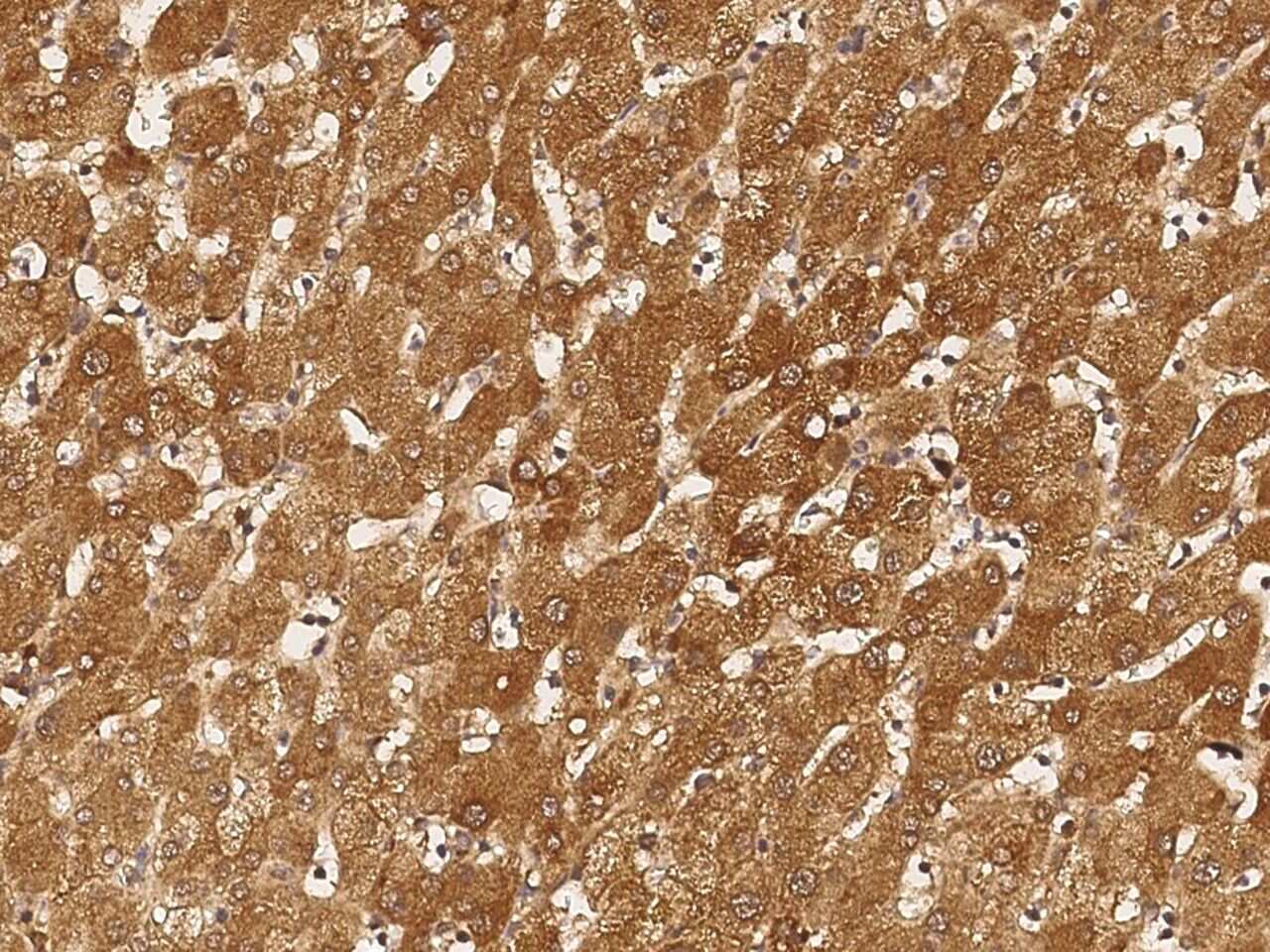
- Immunohistochemical staining of human ACSS3 in human liver with ACSS3 Polyclonal Antibody (Product # PA5-80305, 1:5,000, formalin-fixed paraffin embedded sections).
Supportive validation
- Submitted by
- Invitrogen Antibodies (provider)
- Main image
- Experimental details
- ACSS3 Immunoprecipitation using: Lane A: 0.5 mg HepG2 Whole Cell Lysate 1 µL with ACSS3 Polyclonal Antibody (Product # PA5-80305) and 15 µL of 50 % Protein G agarose. Primary antibody: ACSS3 Polyclonal Antibody, at 1:500 dilution. Secondary antibody: Dylight 800-labeled antibody to rabbit IgG (H+L), at 1:5,000 dilution. Developed using the Odyssey technique. Performed under reducing conditions. Predicted band size: 75 kDa. Observed band size: 75 kDa.

- Submitted by
- Invitrogen Antibodies (provider)
- Main image

- Experimental details
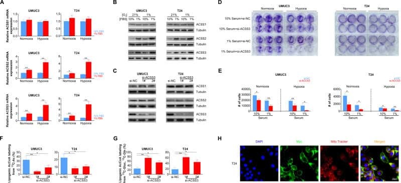
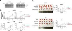
- Fig. 2 ACSS3 is induced under metabolic stress and facilitate BLCA cell growth. a ACSS1, ACSS2, and ACSS3 mRNA expression levels in BLCA cells under normoxia (21% O 2 ) or hypoxia (1% O 2 ) culture with 10% or 1% serum levels. b ACSS1, ACSS2, and ACSS3 protein expression levels in BLCA cells under normoxia (21% O 2 ) or hypoxia (1% O 2 ) culture with 10% or 1% serum levels. c ACSS1, ACSS2, and ACSS3 protein expression levels in BLCA cells transfected with control of ACSS3 siRNA. d Colony formed from BLCA cells transfected with control of ACSS3 siRNA under indicated culture situations. e Cell number of BLCA cells transfected with control of ACSS3 siRNA under indicated culture situations after 48 h incubation. f Percentage of lipogenic AcCoA generation from acetate (labeled by 13 C) in BLCA cells transfected with control of ACSS3 siRNA under hypoxia (1% O 2 ) culture. g Percentage of lipogenic AcCoA generation from glutamine or glucose (labeled by 13 C) in BLCA cells transfected with control of ACSS3 siRNA under hypoxia (1% O 2 ) culture. h Subcellular localization of ACSS3-Myc expressed in T24 cells. ACSS3 was detected with anti-myc antibody. Mitochondria was stained with MitoTracker.
- Submitted by
- Invitrogen Antibodies (provider)
- Main image
- Experimental details
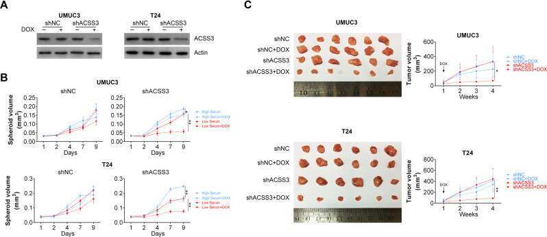
- Fig. 4 ACSS3 is required for spheroid and xenografts formation. a Induction of ACSS3 knockdown by doxycycline in BLCA cells. b Spheroid formation in BLCA cells stably transfected with control of ACSS3 shRNA with or without doxycycline administration under high serum or low serum culture. c Xenografts formed by BLCA cells stably transfected with control of ACSS3 shRNA with or without doxycycline administration under high serum or low serum culture. Right panels are measurements of tumor volumes at indicated time points.
- Submitted by
- Invitrogen Antibodies (provider)
- Main image
- Experimental details
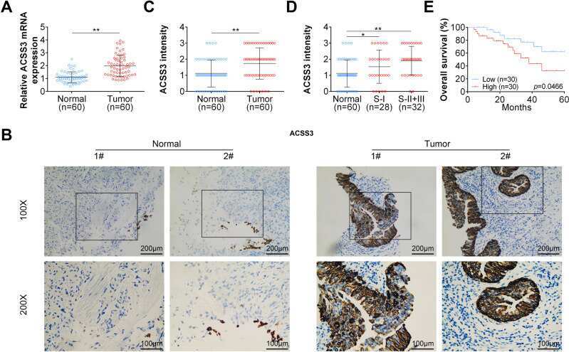
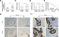
- Fig. 5 ACSS3 is elevated in BLCA patient samples. a ACSS3 mRNA expression levels in BLCA patient samples and adjacent normal tissues. b Representative immunohistochemistry images of ACSS3 staining in BLCA patient samples and adjacent normal tissues. c ACSS3 protein intensities in BLCA patient samples and adjacent normal tissues. d ACSS3 protein intensities in stage I or stage II + III BLCA patient samples and adjacent normal tissues. e Overall survival of BLCA patients with higher or low ACSS3 mRNA expression levels.