Explore
 Validate
 Learn
Learn Western blot
 Immunocytochemistry
Antibody data
- Antibody Data
- Antigen structure
- References [0]
- Comments [0]
- Validations
- Immunocytochemistry [1]
- Chromatin Immunoprecipitation [1]
Submit
Validation data
Reference
Comment
Report error
- Product number
- HPA037926 - Provider product page

- Provider
- Atlas Antibodies
- Proper citation
- Atlas Antibodies Cat#HPA037926, RRID:AB_10672400
- Product name
- Anti-POLR3A
- Antibody type
- Polyclonal
- Description
- Polyclonal Antibody against Human POLR3A, Gene description: polymerase (RNA) III (DNA directed) polypeptide A, 155kDa, Alternative Gene Names: hRPC155, RPC1, RPC155, Validated applications: ChIP, ICC, WB, Uniprot ID: O14802, Storage: Store at +4°C for short term storage. Long time storage is recommended at -20°C.
- Reactivity
- Human
- Host
- Rabbit
- Conjugate
- Unconjugated
- Isotype
- IgG
- Vial size
- 100 µl
- Concentration
- 0.2 mg/ml
- Storage
- Store at +4°C for short term storage. Long time storage is recommended at -20°C.
- Handling
- The antibody solution should be gently mixed before use.
No comments: Submit comment
Supportive validation
- Submitted by
- Atlas Antibodies (provider)
- Main image
- Experimental details
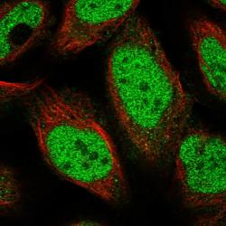
- Immunofluorescent staining of human cell line U-2 OS shows localization to nucleoplasm.
- Sample type
- Human

Supportive validation
- Submitted by
- Atlas Antibodies (provider)
- Main image
- Experimental details
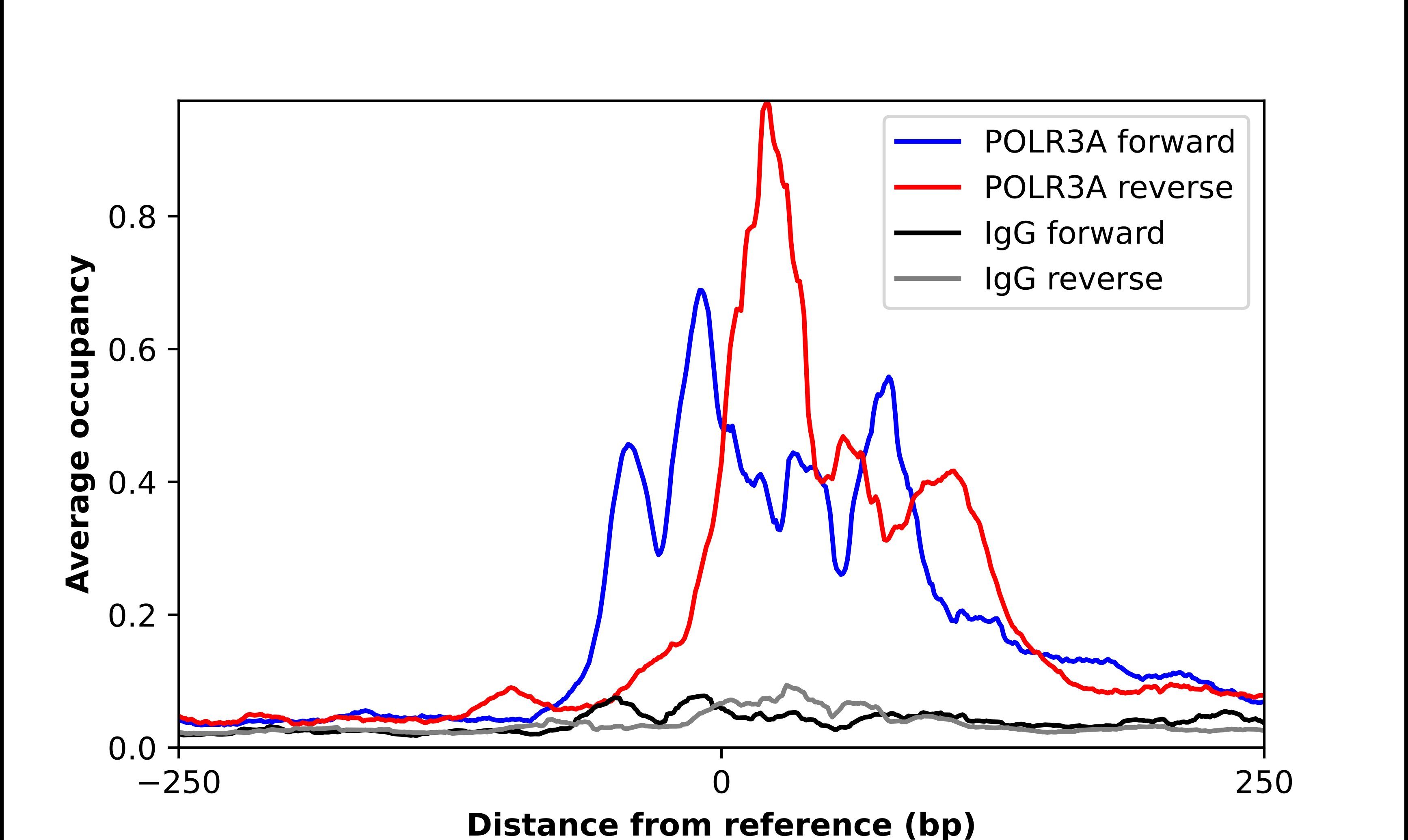
- ChIP-Exo-Seq composite graph for Anti-POLR3A (HPA037926, Lot A114684) tested in K562 cells. Strand-specific reads (blue: forward, red: reverse) and IgG controls (black: forward, grey: reverse) are plotted against the distance from a composite set of reference binding sites. The antibody exhibits robust target enrichment compared to a non-specific IgG control and precisely reveals its structural organization around the binding site. Data generated by Prof. B. F. Pugh´s Lab at Cornell University.