Explore
 Validate
 Learn
Learn Western blot
 Immunocytochemistry
Antibody data
- Antibody Data
- Antigen structure
- References [1]
- Comments [0]
- Validations
- Western blot [4]
- Immunohistochemistry [1]
Submit
Validation data
Reference
Comment
Report error
- Product number
- NBP2-15079 - Provider product page

- Provider
- Novus Biologicals
- Product name
- Rabbit Polyclonal Cyclophilin-F Antibody
- Antibody type
- Polyclonal
- Description
- Immunogen affinity purified.
- Reactivity
- Human, Mouse, Rat
- Host
- Rabbit
- Isotype
- IgG
- Vial size
- 0.1 ml
- Storage
- Aliquot and store at -20C or -80C. Avoid freeze-thaw cycles.
Submitted references Mitochondria Permeability Transition versus Necroptosis in Oxalate-Induced AKI.
Mulay SR, Honarpisheh MM, Foresto-Neto O, Shi C, Desai J, Zhao ZB, Marschner JA, Popper B, Buhl EM, Boor P, Linkermann A, Liapis H, Bilyy R, Herrmann M, Romagnani P, Belevich I, Jokitalo E, Becker JU, Anders HJ
Journal of the American Society of Nephrology : JASN 2019 Oct;30(10):1857-1869
Journal of the American Society of Nephrology : JASN 2019 Oct;30(10):1857-1869
No comments: Submit comment
Supportive validation
- Submitted by
- Novus Biologicals (provider)
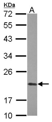
- Main image

- Experimental details
- Western Blot: Cyclophilin-F Antibody [NBP2-15079] - Sample (30 ug of whole cell lysate) A: A431 12% SDS PAGE; antibody diluted at 1:1000.
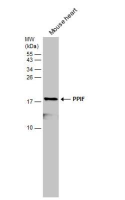
- Submitted by
- Novus Biologicals (provider)
- Main image

- Experimental details
- Western Blot: Cyclophilin-F Antibody [NBP2-15079] - ouse tissue extract (50 ug) was separated by 15% SDS-PAGE, and the membrane was blotted with PPIF antibody [N1C3] diluted at 1:5000.
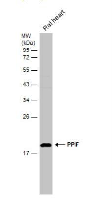
- Submitted by
- Novus Biologicals (provider)
- Main image

- Experimental details
- Western Blot: Cyclophilin-F Antibody [NBP2-15079] - Rat tissue extract (50 ug) was separated by 12% SDS-PAGE, and the membrane was blotted with PPIF antibody [N1C3] diluted at 1:500.
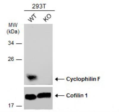
- Submitted by
- Novus Biologicals (provider)
- Main image

- Experimental details
- Western Blot: Cyclophilin-F Antibody [NBP2-15079] - Wild-type (WT) and Cyclophilin F knockout (KO) 293T cell extracts (30 ug) were separated by 12% SDS-PAGE, and the membrane was blotted with Cyclophilin-F antibody diluted at 1:5000. HRP-conjugated anti-rabbit IgG antibody was used to detect the primary antibody.
Supportive validation
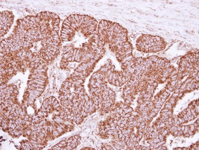
- Submitted by
- Novus Biologicals (provider)
- Main image
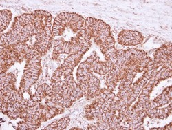
- Experimental details
- Immunohistochemistry-Paraffin: Cyclophilin-F Antibody [NBP2-15079] - Paraffin-embedded Colon ca, using antibody at 1:250 dilution.