 Explore
 Validate
 Learn
Learn Western blot
 Immunocytochemistry
Antibody data
- Antibody Data
- Antigen structure
- References [0]
- Comments [0]
- Validations
- Western blot [2]
- Immunocytochemistry [2]
- Immunohistochemistry [2]
Submit
Validation data
Reference
Comment
Report error
- Product number
- LS-B10822 - Provider product page

- Provider
- LSBio
- Product name
- IHC-plus™ GLO1 / Glyoxalase I Antibody (aa1-184) LS-B10822
- Antibody type
- Polyclonal
- Description
- Immunoaffinity purified
- Reactivity
- Human, Mouse
- Host
- Rabbit
- Isotype
- IgG
- Storage
- Keep as concentrated solution. Aliquot and store at -20°C or below. Avoid multiple freeze-thaw cycles.
No comments: Submit comment
Enhanced validation
- Submitted by
- LSBio (provider)
- Enhanced method
- Genetic validation
- Main image
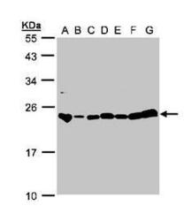
- Experimental details
- Sample (30g whole cell lysate). A:293T, B: A431 , C: H1299, D: HeLa S3 , E: Hep G2 . F: MOLT4 . G: Raji . 12% SDS PAGE. GLO1 / Glyoxalase I antibody diluted at 1:1000

- Submitted by
- LSBio (provider)
- Enhanced method
- Genetic validation
- Main image

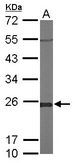
- Experimental details
- Sample (50 ug of whole cell lysate). A: Mouse brain. 12% SDS PAGE. GLO1 / Glyoxalase I antibody diluted at 1:1000.
Supportive validation
- Submitted by
- LSBio (provider)
- Enhanced method
- Genetic validation
- Main image
- Experimental details
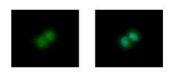
- Glyoxalase I antibody [N1C3] detects GLO1 protein at cytoplasm and nucleus by immunofluorescent analysis. HepG2 cells were fixed in 4% paraformaldehyde at RT for 15 min. GLO1 protein stained by Glyoxalase I antibody [N1C3] diluted at 1:500.

- Submitted by
- LSBio (provider)
- Main image

- Experimental details
- Glyoxalase I antibody [N1C3] detects GLO1 protein at cytoplasm and nucleus by immunofluorescent analysis. HepG2 cells were fixed in 4% paraformaldehyde at RT for 15 min. GLO1 protein stained by Glyoxalase I antibody [N1C3] diluted at 1:500.

Supportive validation
- Submitted by
- LSBio (provider)
- Enhanced method
- Genetic validation
- Main image

- Experimental details
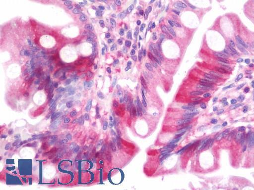
- Anti-GLO1 / Glyoxalase I antibody IHC staining of human small intestine. Immunohistochemistry of formalin-fixed, paraffin-embedded tissue after heat-induced antigen retrieval. Antibody concentration 7.5 ug/ml.
- Submitted by
- LSBio (provider)
- Enhanced method
- Genetic validation
- Main image

- Experimental details
- Anti-GLO1 / Glyoxalase I antibody IHC staining of human small intestine. Immunohistochemistry of formalin-fixed, paraffin-embedded tissue after heat-induced antigen retrieval. Antibody concentration 7.5 ug/ml.
