Explore
 Validate
 Learn
Learn Flow cytometry
Antibody data
- Antibody Data
- Antigen structure
- References [5]
- Comments [0]
- Validations
- Flow cytometry [2]
- Other assay [1]
Submit
Validation data
Reference
Comment
Report error
- Product number
- 12-0792-41 - Provider product page

- Provider
- Invitrogen Antibodies
- Product name
- CD79a Monoclonal Antibody (HM47), PE, eBioscience™
- Antibody type
- Monoclonal
- Antigen
- Other
- Description
- Description: The monoclonal antibody HM47 recognizes the cytoplasmic domain of CD79a, also known as mb-1. CD79a is a 47 kDa membrane glycoprotein that associates with CD79b to form the heterodimeric B cell receptor (BCR). This receptor is responsible for B cell signaling, resulting in activation, apoptosis or anergy. Expression of CD79a is found throughout development from the earliest pre-B cell to plasma cells. CD79 is expressed on B cell neoplasms and some non-B cell malignancies such as AML. Unlike other antibodies that can be masked in B cell neoplastic cells, HM47 is a reliable antibody for recognizing CD79a and, as such, is a good marker for B cells. Because several variants of CD79a exist, the exact location of the epitope may differ slightly. Nevertheless, in humans thie epitope is typically amino acids 208-222. The HM47 antibody reacts to human, mouse, rat, dog, primates (rhesus, chimpanzee, macaque and baboon) pig, guinea rabbit, pig, horse, cow and chicken. Applications Reported: This HM47 antibody has been reported for use in intracellular staining followed by flow cytometric analysis. Applications Tested: This HM47 antibody has been pre-titrated and tested by intracellular staining and flow cytometric analysis of normal human peripheral blood cells. This can be used at 5 µL (0.06 µg) per test. A test is defined as the amount (µg) of antibody that will stain a cell sample in a final volume of 100 µL. Cell number should be determined empirically but can range from 10^5 to 10^8 cells/test. Excitation: 488-561 nm; Emission: 578 nm; Laser: Blue Laser, Green Laser, Yellow-Green Laser. Filtration: 0.2 µm post-manufacturing filtered.
- Reactivity
- Human, Mouse, Rat, Bovine, Canine, Chicken/Avian, Guinea Pig, Porcine
- Host
- Mouse
- Conjugate
- Yellow dye
- Isotype
- IgG
- Antibody clone number
- HM47
- Vial size
- 25 Tests
- Concentration
- 5 μL/Test
- Storage
- 4°C, store in dark, DO NOT FREEZE!
Submitted references In Vitro Expansion and Characterization of Mesenchymal Stromal Cells from Peritoneal Dialysis Effluent in a Human Protein Medium.
Long-term spironolactone treatment reduces coronary TRPC expression, vasoconstriction, and atherosclerosis in metabolic syndrome pigs.
The IgM receptor FcμR limits tonic BCR signaling by regulating expression of the IgM BCR.
Human umbilical cord mesenchymal stem cells improve the reserve function of perimenopausal ovary via a paracrine mechanism.
Differential expression of B29 (CD79b) and mb-1 (CD79a) proteins in acute lymphoblastic leukaemia.
Han B, Zhou L, Guan Q, da Roza G, Wang H, Du C
Stem cells international 2018;2018:5868745
Stem cells international 2018;2018:5868745
Long-term spironolactone treatment reduces coronary TRPC expression, vasoconstriction, and atherosclerosis in metabolic syndrome pigs.
Li W, Chen X, Riley AM, Hiett SC, Temm CJ, Beli E, Long X, Chakraborty S, Alloosh M, White FA, Grant MB, Sturek M, Obukhov AG
Basic research in cardiology 2017 Sep;112(5):54
Basic research in cardiology 2017 Sep;112(5):54
The IgM receptor FcμR limits tonic BCR signaling by regulating expression of the IgM BCR.
Nguyen TT, Kläsener K, Zürn C, Castillo PA, Brust-Mascher I, Imai DM, Bevins CL, Reardon C, Reth M, Baumgarth N
Nature immunology 2017 Mar;18(3):321-333
Nature immunology 2017 Mar;18(3):321-333
Human umbilical cord mesenchymal stem cells improve the reserve function of perimenopausal ovary via a paracrine mechanism.
Li J, Mao Q, He J, She H, Zhang Z, Yin C
Stem cell research & therapy 2017 Mar 9;8(1):55
Stem cell research & therapy 2017 Mar 9;8(1):55
Differential expression of B29 (CD79b) and mb-1 (CD79a) proteins in acute lymphoblastic leukaemia.
Astsaturov IA, Matutes E, Morilla R, Seon BK, Mason DY, Farahat N, Catovsky D
Leukemia 1996 May;10(5):769-73
Leukemia 1996 May;10(5):769-73
No comments: Submit comment
Supportive validation
- Submitted by
- Invitrogen Antibodies (provider)
- Main image
- Experimental details
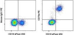
- Surface staining of normal human peripheral blood cells with Anti-Human CD19 eFluor® 450 (Product # 48-0199-42) followed by intraceullular staining with Mouse IgG1 K Isotype Control PE (Product # 12-4714-81) (left) or Anti-Human CD79a PE (right) using IC Fixation and Permeabilization Buffers. Cells in the lymphocyte gate were used for analysis.
- Conjugate
- Yellow dye

- Submitted by
- Invitrogen Antibodies (provider)
- Main image
- Experimental details
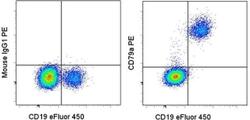
- Surface staining of normal human peripheral blood cells with Anti-Human CD19 eFluor® 450 (Product # 48-0199-42) followed by intraceullular staining with Mouse IgG1 K Isotype Control PE (Product # 12-4714-81) (left) or Anti-Human CD79a PE (right) using IC Fixation and Permeabilization Buffers. Cells in the lymphocyte gate were used for analysis.

Supportive validation
- Submitted by
- Invitrogen Antibodies (provider)
- Main image

- Experimental details
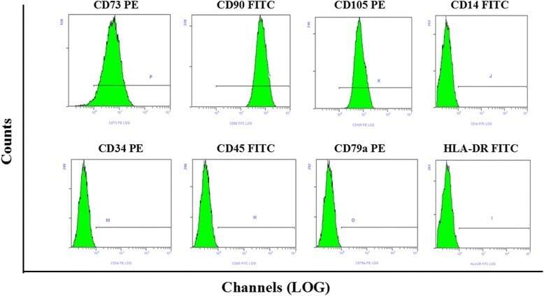
- Fig. 1 Flow cytometry analysis of phenotype characterization of hUCMSCs. Phenotype of CD73, CD90, CD105, CD14, CD34, CD45, CD79a and HLA-DR of hUCMSCs was detected by flow cytometry. Intensity >= 95% represented strong expression while
- Conjugate
- Yellow dye