Explore
 Validate
 Learn
Learn Flow cytometry
Antibody data
- Antibody Data
- Antigen structure
- References [4]
- Comments [0]
- Validations
- Flow cytometry [2]
Submit
Validation data
Reference
Comment
Report error
- Product number
- 47-0792-41 - Provider product page

- Provider
- Invitrogen Antibodies
- Product name
- Anti-CD79a Monoclonal Antibody (HM47), APC-eFluor 780, eBioscience™
- Antibody type
- Monoclonal
- Antigen
- Other
- Description
- Description: The monoclonal antibody HM47 recognizes the cytoplasmic domain of CD79a, also known as mb-1. CD79a is a 47 kDa membrane glycoprotein that associates with CD79b to form the heterodimeric B cell receptor (BCR). This receptor is responsible for B cell signaling, resulting in activation, apoptosis or anergy. Expression of CD79a is found throughout development from the earliest pre-B cell to plasma cells. CD79 is expressed on B cell neoplasms and some non-B cell malignancies such as AML. Unlike other antibodies that can be masked in B cell neoplastic cells, HM47 is a reliable antibody for recognizing CD79a and, as such, is a good marker for B cells. Because several variants of CD79a exist, the exact location of the epitope may differ slightly. Nevertheless, in humans thie epitope is typically amino acids 208-222. The HM47 antibody reacts to human, mouse, rat, dog, primates (rhesus, chimpanzee, macaque and baboon) pig, guinea rabbit, pig, horse, cow and chicken. Applications Reported: This HM47 antibody has been reported for use in intracellular staining followed by flow cytometric analysis. Applications Tested: This HM47 antibody has been pre-titrated and tested by intracelµLlar staining and flow cytometric analysis of normal human peripheral blood cells. This can be used at 5 µL (0.06 µg) per test. A test is defined as the amount (µg) of antibody that will stain a cell sample in a final volume of 100 µL. Cell number should be determined empirically but can range from 10^5 to 10^8 cells/test. APC-eFluor 780 emits at 780 nm and is excited with the Red laser (633 nm). Please make sure that your instrument is capable of detecting this fluorochome. Light sensitivity: This tandem is sensitive to photo-induced oxidation. Please protect this vial and stained samples from light. Fixation: Samples can be stored in IC Fixation Buffer (cat. 00-8222) (100 µL cell sample + 100 µL IC Fixation Buffer) or 1-step Fix/Lyse Solution (cat. 00-5333) for up to 3 days in the dark at 4°C with minimal impact on brightness and FRET efficiency/compensation. Some generalizations regarding fluorophore performance after fixation can be made, but clone specific performance should be determined empirically. Excitation: 633-647 nm; Emission: 780 nm; Laser: Red Laser. Filtration: 0.2 µm post-manufacturing filtered.
- Reactivity
- Human, Mouse, Rat, Bovine, Canine, Chicken/Avian, Guinea Pig, Porcine
- Host
- Mouse
- Isotype
- IgG
- Antibody clone number
- HM47
- Vial size
- 25 Tests
- Concentration
- 5 µL/Test
- Storage
- 4° C, store in dark, DO NOT FREEZE!
Submitted references Human umbilical cord mesenchymal stem cells improve the reserve function of perimenopausal ovary via a paracrine mechanism.
Antiviral antibodies are necessary for control of simian immunodeficiency virus replication.
CD79a is heterogeneously expressed in neoplastic and normal myeloid precursors and megakaryocytes in an antibody clone-dependent manner.
Differential expression of B29 (CD79b) and mb-1 (CD79a) proteins in acute lymphoblastic leukaemia.
Li J, Mao Q, He J, She H, Zhang Z, Yin C
Stem cell research & therapy 2017 Mar 9;8(1):55
Stem cell research & therapy 2017 Mar 9;8(1):55
Antiviral antibodies are necessary for control of simian immunodeficiency virus replication.
Miller CJ, Genescà M, Abel K, Montefiori D, Forthal D, Bost K, Li J, Favre D, McCune JM
Journal of virology 2007 May;81(10):5024-35
Journal of virology 2007 May;81(10):5024-35
CD79a is heterogeneously expressed in neoplastic and normal myeloid precursors and megakaryocytes in an antibody clone-dependent manner.
Bhargava P, Kallakury BV, Ross JS, Azumi N, Bagg A
American journal of clinical pathology 2007 Aug;128(2):306-13
American journal of clinical pathology 2007 Aug;128(2):306-13
Differential expression of B29 (CD79b) and mb-1 (CD79a) proteins in acute lymphoblastic leukaemia.
Astsaturov IA, Matutes E, Morilla R, Seon BK, Mason DY, Farahat N, Catovsky D
Leukemia 1996 May;10(5):769-73
Leukemia 1996 May;10(5):769-73
No comments: Submit comment
Supportive validation
- Submitted by
- Invitrogen Antibodies (provider)
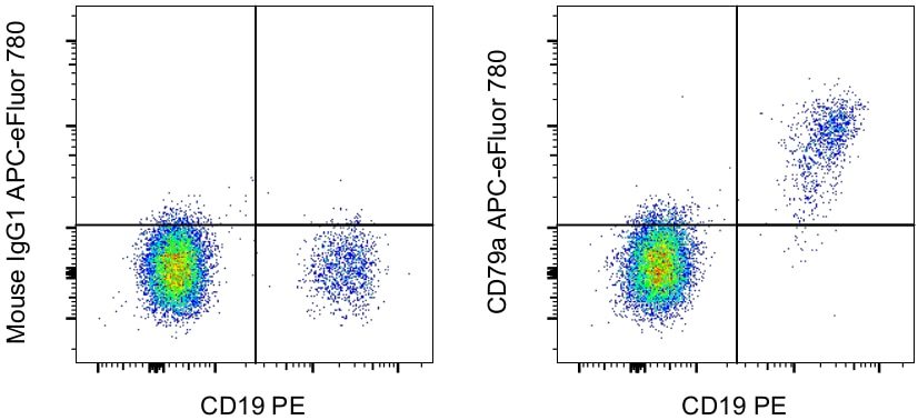
- Main image

- Experimental details
- Intracellular staining of normal human peripheral blood cells with Anti-Human CD19 PE (Product # 12-0199-80) and Mouse IgG1 K Isotype Control APC-eFluor® 780 (Product # 47-4714-82) (left) or Anti-CD79a APC-eFluor® 780 (right). Cells in the lymphocyte gate were used for analysis.
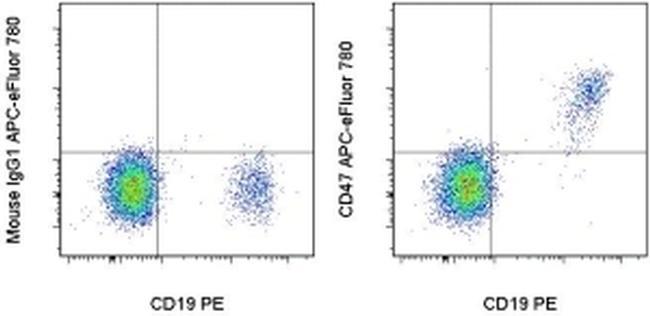
- Submitted by
- Invitrogen Antibodies (provider)
- Main image

- Experimental details
- Intracellular staining of normal human peripheral blood cells with Anti-Human CD19 PE (Product # 12-0199-80) and Mouse IgG1 K Isotype Control APC-eFluor® 780 (Product # 47-4714-82) (left) or Anti-CD79a APC-eFluor® 780 (right). Cells in the lymphocyte gate were used for analysis.