Explore
 Validate
 Learn
Learn Western blot
Antibody data
- Antibody Data
- Antigen structure
- References [5]
- Comments [0]
- Validations
- Western blot [1]
- Immunohistochemistry [1]
- Flow cytometry [3]
Submit
Validation data
Reference
Comment
Report error
- Product number
- MA5-11636 - Provider product page

- Provider
- Invitrogen Antibodies
- Product name
- Anti-CD79a Monoclonal Antibody (JCB117)
- Antibody type
- Monoclonal
- Antigen
- Recombinant full-length protein
- Description
- MA5-11636 targets CD79a in WB, FACS and IHC (P) applications and shows reactivity with Human and Mouse samples. This antibody is not suitable for mouse BAF-3 cell lysates in Western blot analysis. This antibody also detects some non-specific bands on Ramos cell lysates. The MA5-11636 immunogen is recombinant protein containing part of the extracellular portion of the CD79a glycoprotein.
- Reactivity
- Human, Mouse
- Host
- Mouse
- Isotype
- IgG
- Antibody clone number
- JCB117
- Vial size
- 500 µL
- Concentration
- 0.2 mg/mL
- Storage
- 4° C
Submitted references Structural and functional studies of Igalphabeta and its assembly with the B cell antigen receptor.
Hyperplasia of hair follicles and other adnexal structures in cutaneous lymphoproliferative disorders: a study of 53 cases, including so-called pseudolymphomatous folliculitis and overt lymphomas.
Primary cutaneous histiocyte and neutrophil-rich CD30+ and CD56+ anaplastic large-cell lymphoma with prominent angioinvasion and nerve involvement in the forehead and scalp of an immunocompetent woman.
Cutaneous lymphoid hyperplasia and other lymphoid infiltrates of the breast nipple: a retrospective clinicopathologic study of fifty-six patients.
Cutaneous lymphoid hyperplasia and other lymphoid infiltrates of the breast nipple: a retrospective clinicopathologic study of fifty-six patients.
Radaev S, Zou Z, Tolar P, Nguyen K, Nguyen A, Krueger PD, Stutzman N, Pierce S, Sun PD
Structure (London, England : 1993) 2010 Aug 11;18(8):934-43
Structure (London, England : 1993) 2010 Aug 11;18(8):934-43
Hyperplasia of hair follicles and other adnexal structures in cutaneous lymphoproliferative disorders: a study of 53 cases, including so-called pseudolymphomatous folliculitis and overt lymphomas.
Kazakov DV, Belousova IE, Kacerovska D, Sima R, Vanecek T, Vazmitel M, Pizinger K, Michal M
The American journal of surgical pathology 2008 Oct;32(10):1468-78
The American journal of surgical pathology 2008 Oct;32(10):1468-78
Primary cutaneous histiocyte and neutrophil-rich CD30+ and CD56+ anaplastic large-cell lymphoma with prominent angioinvasion and nerve involvement in the forehead and scalp of an immunocompetent woman.
Boudova L, Kazakov DV, Jindra P, Sima R, Vanecek T, Kuntscher V, Vera V, Bouda J, Michal M
Journal of cutaneous pathology 2006 Aug;33(8):584-9
Journal of cutaneous pathology 2006 Aug;33(8):584-9
Cutaneous lymphoid hyperplasia and other lymphoid infiltrates of the breast nipple: a retrospective clinicopathologic study of fifty-six patients.
Boudova L, Kazakov DV, Sima R, Vanecek T, Torlakovic E, Lamovec J, Kutzner H, Szepe P, Plank L, Bouda J, Hes O, Mukensnabl P, Michal M
The American Journal of dermatopathology 2005 Oct;27(5):375-86
The American Journal of dermatopathology 2005 Oct;27(5):375-86
No comments: Submit comment
Supportive validation
- Submitted by
- Invitrogen Antibodies (provider)
- Main image
- Experimental details
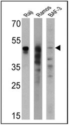
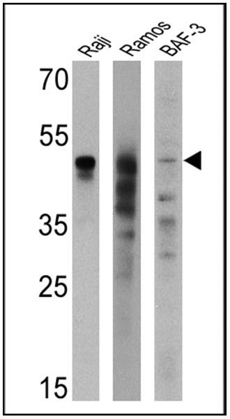
- Western blot analysis of CD79a was performed by loading 25 µg of Raji (Lane 1), Ramos (Lane 2), and BAF-3 cell lysates (Lane 3) and a molecular weight protein ladder onto an SDS polyacrylamide gel. Proteins were transferred to a PVDF membrane and blocked with a blocking buffer at 4ºC overnight. The membrane was probed with a CD79a monoclonal antibody (Product # MA5-11636) at a dilution of 1:200 (Raji and Ramos) and 1:100 (BAF-3) overnight at 4°C, washed in TBST, and probed with an HRP-conjugated secondary antibody for 1 hr at room temperature in the dark. Chemiluminescent detection was performed using Pierce ECL Plus Western Blotting Substrate (Product # 32132). Results show a band at 47 kDa in Raji and Ramos cell lines.
Supportive validation
- Submitted by
- Invitrogen Antibodies (provider)
- Main image

- Experimental details
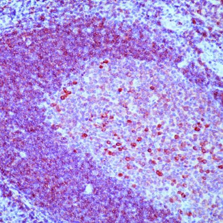
- Formalin-fixed, paraffin-embedded human tonsil stained with CD79a antibody using peroxidase-conjugate and AEC chromogen. Note membrane staining of B cells.
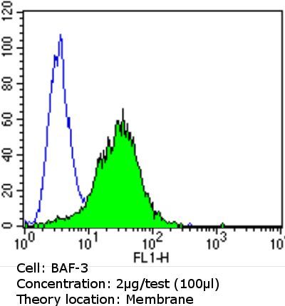
Supportive validation
- Submitted by
- Invitrogen Antibodies (provider)
- Main image
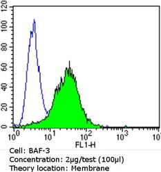
- Experimental details
- Flow cytometry analysis of CD79a in BAF-3 cells compared to an isotype control (blue). Cells were harvested, adjusted to a concentration of 1-5x10^6 cells/mL, fixed with 2% paraformaldehyde and washed with PBS. Cells were blocked with a 2% solution of BSA-PBS for 30 min at room temperature and incubated with a CD79a monoclonal antibody (Product # MA5-11636) at a dilution of 2 µg/test for 60 min at room temperature. Cells were then incubated for 40 min at room temperature in the dark using a Dylight 488-conjugated goat anti-mouse IgG (H+L) secondary antibody and re-suspended in PBS for FACS analysis.
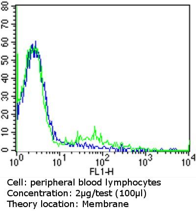
- Submitted by
- Invitrogen Antibodies (provider)
- Main image
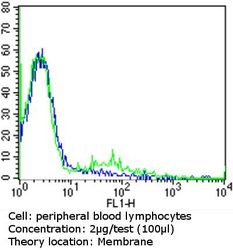
- Experimental details
- Flow cytometry analysis of CD79a in peripheral blood mononuclear cells compared to an isotype control (blue). Human blood was collected, combined with a hydrophilic polysaccharide, centrifuged, transferred to a conical tube and washed with PBS. 50 µL of cell solution was added to each tube at a dilution of 2x10^7 cells/mL, followed by the addition of 50 µL of isotype control and primary antibody (Product # MA5-11636) at a dilution of 2 µg/test. Cells were incubated for 30 min at 4ºC and washed with a cell buffer, followed by incubation with a DyLight 488-conjugated goat anti-mouse IgG (H+L) secondary for 30 min at 4ºC in the dark. FACS analysis was performed using 400 µL of cell buffer.
- Submitted by
- Invitrogen Antibodies (provider)
- Main image
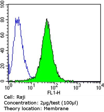
- Experimental details
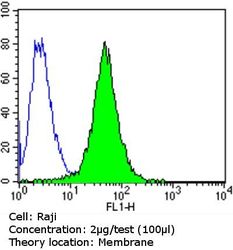
- Flow cytometry analysis of CD79a in Raji cells compared to an isotype control (blue). Cells were harvested, adjusted to a concentration of 1-5x10^6 cells/mL, fixed with 2% paraformaldehyde and washed with PBS. Cells were blocked with a 2% solution of BSA-PBS for 30 min at room temperature and incubated with a CD79a monoclonal antibody (Product # MA5-11636) at a dilution of 2 µg/test for 60 min at room temperature. Cells were then incubated for 40 min at room temperature in the dark using a Dylight 488-conjugated goat anti-mouse IgG (H+L) secondary antibody and re-suspended in PBS for FACS analysis.