Explore
 Validate
 Learn
Learn Western blot
Antibody data
- Antibody Data
- Antigen structure
- References [1]
- Comments [0]
- Validations
- Western blot [1]
- Immunohistochemistry [1]
Submit
Validation data
Reference
Comment
Report error
- Product number
- AF7768 - Provider product page

- Provider
- R&D Systems
- Product name
- Human MEGF9 Antibody
- Antibody type
- Polyclonal
- Description
- Immunogen affinity purified. Detects human MEGF9 in direct ELISAs and Western blots.
- Reactivity
- Human
- Host
- Sheep
- Conjugate
- Unconjugated
- Antigen sequence
Q9H1U4- Isotype
- IgG
- Vial size
- 100 ug
- Concentration
- LYOPH
- Storage
- Use a manual defrost freezer and avoid repeated freeze-thaw cycles. 12 months from date of receipt, -20 to -70 °C as supplied. 1 month, 2 to 8 °C under sterile conditions after reconstitution. 6 months, -20 to -70 °C under sterile conditions after reconstitution.
Submitted references Functionally distinct PI 3-kinase pathways regulate myelination in the peripheral nervous system.
Heller BA, Ghidinelli M, Voelkl J, Einheber S, Smith R, Grund E, Morahan G, Chandler D, Kalaydjieva L, Giancotti F, King RH, Fejes-Toth AN, Fejes-Toth G, Feltri ML, Lang F, Salzer JL
The Journal of cell biology 2014 Mar 31;204(7):1219-36
The Journal of cell biology 2014 Mar 31;204(7):1219-36
No comments: Submit comment
Supportive validation
- Submitted by
- R&D Systems (provider)
- Main image
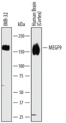
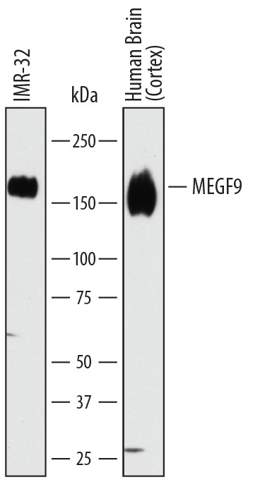
- Experimental details
- Detection of Human MEGF9 by Western Blot. Western blot shows lysates of IMR-32 human neuroblastoma cell line and human brain (cortex) tissue. PVDF membrane was probed with 1 µg/mL of Sheep Anti-Human MEGF9 Antigen Affinity-purified Polyclonal Antibody (Catalog # AF7768) followed by HRP-conjugated Anti-Sheep IgG Secondary Antibody (Catalog # HAF016). A specific band was detected for MEGF9 at approximately 160 kDa (as indicated). This experiment was conducted under reducing conditions and using Immunoblot Buffer Group 1.
Supportive validation
- Submitted by
- R&D Systems (provider)
- Main image
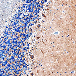
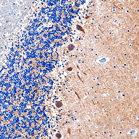
- Experimental details
- MEGF9 in Human Brain. MEGF9 was detected in immersion fixed paraffin-embedded sections of human brain (cerebellum) using Sheep Anti-Human MEGF9 Antigen Affinity-purified Polyclonal Antibody (Catalog # AF7768) at 1 µg/mL overnight at 4 °C. Before incubation with the primary antibody, tissue was subjected to heat-induced epitope retrieval using Antigen Retrieval Reagent-Basic (Catalog # CTS013). Tissue was stained using the Anti-Sheep HRP-DAB Cell & Tissue Staining Kit (brown; Catalog # CTS019) and counter-stained with hematoxylin (blue). Specific staining was localized to Purkinje neurons. View our protocol for Chromogenic IHC Staining of Paraffin-embedded Tissue Sections.