 Explore
 Validate
 Learn
Learn Western blot
Antibody data
- Antibody Data
- Antigen structure
- References [10]
- Comments [0]
- Validations
- Western blot [5]
- Immunocytochemistry [3]
- Immunohistochemistry [1]
Submit
Validation data
Reference
Comment
Report error
- Product number
- GTX30114 - Provider product page

- Provider
- GeneTex
- Proper citation
- GeneTex Cat#GTX30114, RRID:AB_370290
- Product name
- HIF2 alpha antibody
- Antibody type
- Polyclonal
- Reactivity
- Human, Mouse, Rat, Simian
- Host
- Rabbit
Submitted references Metabolic consequences of HIF silencing in a triple negative human breast cancer xenograft.
Hypoxia inducible factor 2α (HIF2α/EPAS1) is associated with development of pulmonary hypertension in severe congenital diaphragmatic hernia patients.
Prognostic significance of the programmed death ligand 1 expression in clear cell renal cell carcinoma and correlation with the tumor microenvironment and hypoxia-inducible factor expression.
Hypoxia favors myosin heavy chain beta gene expression in an Hif-1alpha-dependent manner.
Melatonin promotes cardiomyogenesis of embryonic stem cells via inhibition of HIF-1α stabilization.
Activation of carbonic anhydrase IX by alternatively spliced tissue factor under late-stage tumor conditions.
HIF1α is necessary for exercise-induced neuroprotection while HIF2α is needed for dopaminergic neuron survival in the substantia nigra pars compacta.
Prolyl-4-hydroxylase 2 enhances hypoxia-induced glioblastoma cell death by regulating the gene expression of hypoxia-inducible factor-α.
Expression of HIF-1alpha by human macrophages: implications for the use of macrophages in hypoxia-regulated cancer gene therapy.
Ubiquitination of hypoxia-inducible factor requires direct binding to the beta-domain of the von Hippel-Lindau protein.
Bharti SK, Mironchik Y, Wildes F, Penet MF, Goggins E, Krishnamachary B, Bhujwalla ZM
Oncotarget 2018 Mar 16;9(20):15326-15339
Oncotarget 2018 Mar 16;9(20):15326-15339
Hypoxia inducible factor 2α (HIF2α/EPAS1) is associated with development of pulmonary hypertension in severe congenital diaphragmatic hernia patients.
Huang Y, Boerema-de Munck A, Buscop-van Kempen M, Sluiter I, de Krijger R, Tibboel D, Rottier RJ
Pulmonary circulation 2018 Jul-Sep;8(3):2045894018783734
Pulmonary circulation 2018 Jul-Sep;8(3):2045894018783734
Prognostic significance of the programmed death ligand 1 expression in clear cell renal cell carcinoma and correlation with the tumor microenvironment and hypoxia-inducible factor expression.
Tatli Dogan H, Kiran M, Bilgin B, Kiliçarslan A, Sendur MAN, Yalçin B, Ardiçoglu A, Atmaca AF, Gumuskaya B
Diagnostic pathology 2018 Aug 25;13(1):60
Diagnostic pathology 2018 Aug 25;13(1):60
Hypoxia favors myosin heavy chain beta gene expression in an Hif-1alpha-dependent manner.
Binó L, Procházková J, Radaszkiewicz KA, Kučera J, Kudová J, Pacherník J, Kubala L
Oncotarget 2017 Oct 13;8(48):83684-83697
Oncotarget 2017 Oct 13;8(48):83684-83697
Melatonin promotes cardiomyogenesis of embryonic stem cells via inhibition of HIF-1α stabilization.
Kudová J, Vašíček O, Číž M, Kubala L
Journal of pineal research 2016 Nov;61(4):493-503
Journal of pineal research 2016 Nov;61(4):493-503
Activation of carbonic anhydrase IX by alternatively spliced tissue factor under late-stage tumor conditions.
Ramchandani D, Unruh D, Lewis CS, Bogdanov VY, Weber GF
Laboratory investigation; a journal of technical methods and pathology 2016 Dec;96(12):1234-1245
Laboratory investigation; a journal of technical methods and pathology 2016 Dec;96(12):1234-1245
HIF1α is necessary for exercise-induced neuroprotection while HIF2α is needed for dopaminergic neuron survival in the substantia nigra pars compacta.
Smeyne M, Sladen P, Jiao Y, Dragatsis I, Smeyne RJ
Neuroscience 2015 Jun 4;295:23-38
Neuroscience 2015 Jun 4;295:23-38
Prolyl-4-hydroxylase 2 enhances hypoxia-induced glioblastoma cell death by regulating the gene expression of hypoxia-inducible factor-α.
Sun W, Jelkmann W, Depping R
Cell death & disease 2014 Jul 10;5:e1322
Cell death & disease 2014 Jul 10;5:e1322
Expression of HIF-1alpha by human macrophages: implications for the use of macrophages in hypoxia-regulated cancer gene therapy.
Burke B, Tang N, Corke KP, Tazzyman D, Ameri K, Wells M, Lewis CE
The Journal of pathology 2002 Feb;196(2):204-12
The Journal of pathology 2002 Feb;196(2):204-12
Ubiquitination of hypoxia-inducible factor requires direct binding to the beta-domain of the von Hippel-Lindau protein.
Ohh M, Park CW, Ivan M, Hoffman MA, Kim TY, Huang LE, Pavletich N, Chau V, Kaelin WG
Nature cell biology 2000 Jul;2(7):423-7
Nature cell biology 2000 Jul;2(7):423-7
No comments: Submit comment
Supportive validation
- Submitted by
- GeneTex (provider)
- Main image

- Experimental details
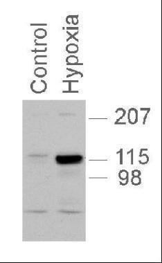
- GTX30114 utilized in western blot with PC12 nuclear extracts, 1:1,000, 5 second exposure.
- Submitted by
- GeneTex (provider)
- Main image
- Experimental details
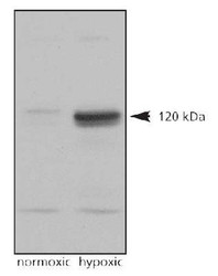
- Western Blot: HIF-2 alpha Antibody (GTX30114) - Analysis on normoxic and hypoxic nuclear rat cell lysates.
- Submitted by
- GeneTex (provider)
- Main image
- Experimental details
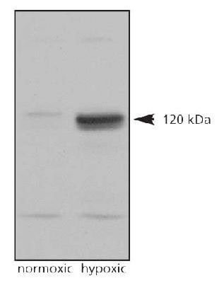
- Western Blot: HIF-2 alpha Antibody (GTX30114) - Analysis of HIF-2 alpha on Lane 1, Cobalt chloride treated COS7 nuclear extracts and Lane 2, Untreated COS7 nuclear extracts using GTX30114

- Submitted by
- GeneTex (provider)
- Main image
- Experimental details
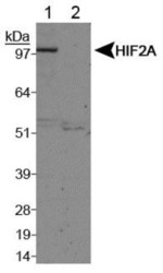
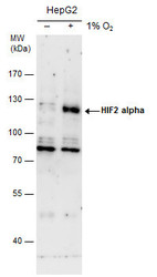
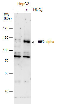
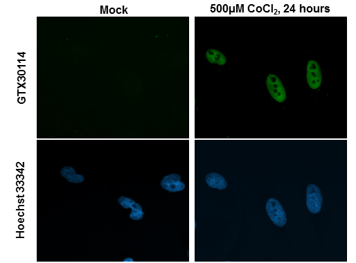
- HIF2 alpha antibody detects HIF2 alpha protein by western blot analysis. Un-treated (-) and treated (+, 0.01 O2 treatment for 24h) HepG2 whole cell extracts (30 ?g) were separated by 7.5% SDS-PAGE, and the membrane was blotted with HIF2 alpha antibody (GTX30114) diluted by 1:500. The HRP-conjugated anti-rabbit IgG antibody (GTX213110-01) was used to detect the primary antibody.
- Submitted by
- GeneTex (provider)
- Main image
- Experimental details
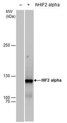
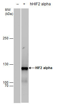
- HIF2 alpha antibody detects HIF2 alpha protein by western blot analysis. Non-transfected (-) and HIF2A-transfected (+, including Myc-DDK-tag) 293T whole cell extracts (30 ?g) were separated by 5% SDS-PAGE, and the membrane was blotted with HIF2 alpha antibody (GTX30114) diluted by 1:1000. The HRP-conjugated anti-rabbit IgG antibody (GTX213110-01) was used to detect the primary antibody.
Supportive validation
- Submitted by
- GeneTex (provider)
- Main image
- Experimental details
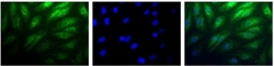
- Immunocytochemistry/Immunofluorescence: HIF-2 alpha Antibody (GTX30114) - Detection of HIF-2 Alpha (Green) in RCC4 cells using GTX30114. Nuclei (Blue) are counterstained with Hoechst 33258.
- Submitted by
- GeneTex (provider)
- Main image
- Experimental details
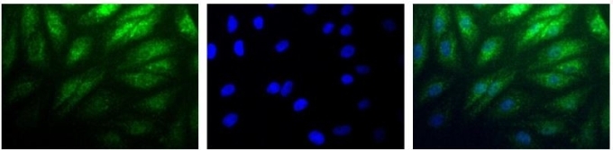
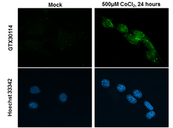
- HIF2 alpha antibody detects HIF2 alpha protein at nucleus by immunofluorescent analysis.Sample: HeLa cells were fixed in 4% paraformaldehyde at RT for 15 min.Green: HIF2 alpha stained by HIF2 alpha antibody (GTX30114) diluted at 1:200.Blue: Hoechst 33342 staining.
- Submitted by
- GeneTex (provider)
- Main image
- Experimental details
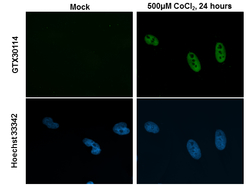
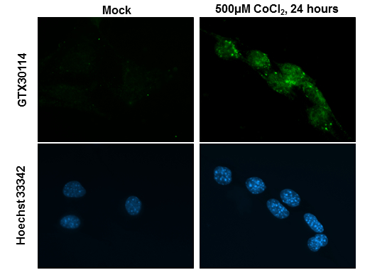
- HIF2 alpha antibody detects HIF2 alpha protein at nucleus by immunofluorescent analysis.Sample: NIH-3T3 cells were fixed in 4% paraformaldehyde at RT for 15 min.Green: HIF2 alpha stained by HIF2 alpha antibody (GTX30114) diluted at 1:200.Blue: Hoechst 33342 staining.
Supportive validation
- Submitted by
- GeneTex (provider)
- Main image
- Experimental details
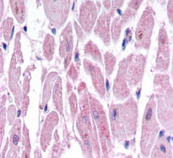
- Immunohistochemistry: HIF-2 alpha Antibody (GTX30114) - Hif-2 alpha immunoreactivity in human cardiac myocytes
