Explore
 Validate
 Learn
Learn Western blot
Antibody data
- Antibody Data
- Antigen structure
- References [0]
- Comments [0]
- Validations
- Western blot [1]
- Immunohistochemistry [1]
Submit
Validation data
Reference
Comment
Report error
- Product number
- PA3-029 - Provider product page

- Provider
- Invitrogen Antibodies
- Product name
- OPRL1 Polyclonal Antibody
- Antibody type
- Polyclonal
- Antigen
- Other
- Description
- Super Bright 436 can be excited with the violet laser line (405 nm) and emits at 436 nm. We recommend using a 450/50 bandpass filter, or equivalent. Please make sure that your instrument is capable of detecting this fluorochrome.
- Reactivity
- Human
- Host
- Rabbit
- Isotype
- IgG
- Vial size
- 100 µL
- Concentration
- Conc. Not Determined
- Storage
- -20°C
No comments: Submit comment
Supportive validation
- Submitted by
- Invitrogen Antibodies (provider)
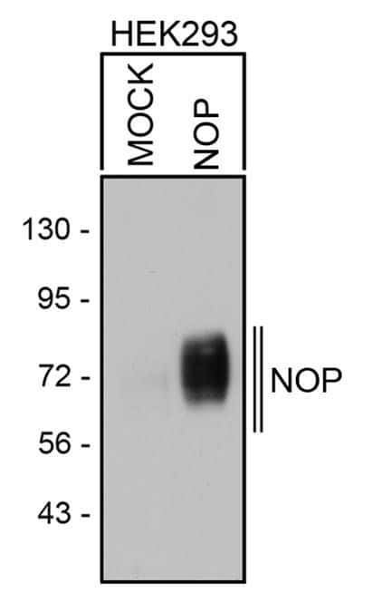
- Main image

- Experimental details
- Western blot analysis of NOP was performed by loading equal amounts of wheat germ lectin agarose bead enriched NOP receptor fractions from mock-transfected or NOP transfected HEK293 lysates onto a 7.5% Tris-HCl polyacrylamide gel. Proteins were transferred to a PVDF membrane, blocked and probed with a NOP polyclonal antibody (Product # PA3-029) at a dilution of 1:5000, overnight at 4C on a rocking platform, followed by an HRP-conjugated goat anti-rabbit IgG secondary antibody. Denatured NOP was detected at ~72kDa. Chemiluminescent detection was performed using ECL.
Supportive validation
- Submitted by
- Invitrogen Antibodies (provider)
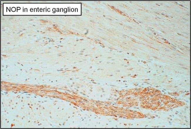
- Main image

- Experimental details
- Immunohistochemistry analysis of NOP was performed on human enteric ganglion tissue. To expose target proteins, antigen retrieval was performed by microwaving tissues for 20 minutes in 10mM sodium citrate buffer (pH 6.0). Tissue slides were probed with a NOP polyclonal antibody (Product # PA3-029) at a dilution of 1:3000, overnight at 4C in a humidified chamber. Tissues were washed, and detection was performed using an ABC kit composed of biotinylated goat anti-rabbit IgG, peroxidase-conjugated avidin, and 3-amino-9-ethylcarbazole (AEC) substrate in acetate buffer. Tissues were counterstained with hematoxylin and dehydrated to prep for mounting.