Explore
 Validate
 Learn
Learn Western blot
 Immunoprecipitation
Antibody data
- Antibody Data
- Antigen structure
- References [4]
- Comments [0]
- Validations
- Western blot [2]
Submit
Validation data
Reference
Comment
Report error
- Product number
- AF1286 - Provider product page

- Provider
- Novus Biologicals
- Product name
- Goat Polyclonal Cystatin E/M/CST6 Antibody
- Antibody type
- Polyclonal
- Description
- Antigen Affinity-purified. Detects human Cystatin E/M in direct ELISAs and Western blots. In direct ELISAs, approximately 25% cross-reactivity with recombinant mouse Cystatin E/M is observed.
- Reactivity
- Human
- Host
- Goat
- Conjugate
- Unconjugated
- Isotype
- IgG
- Vial size
- 100 ug
- Concentration
- LYOPH
- Storage
- Use a manual defrost freezer and avoid repeated freeze-thaw cycles. 12 months from date of receipt, -20 to -70 degreesC as supplied. 1 month, 2 to 8 degreesC under sterile conditions after reconstitution. 6 months, -20 to -70 degreesC under sterile conditions after reconstitution.
Submitted references Glycosylation is important for legumain localization and processing to active forms but not for cystatin E/M inhibitory functions.
Differential secretome analysis reveals CST6 as a suppressor of breast cancer bone metastasis.
Cystatin E/M suppresses legumain activity and invasion of human melanoma.
Invasion suppressor cystatin E/M (CST6): high-level cell type-specific expression in normal brain and epigenetic silencing in gliomas.
Lunde NN, Haugen MH, Bodin Larsen KB, Damgaard I, Pettersen SJ, Kasem R, Rut W, Drag M, Poreba M, Johansen HT, Solberg R
Biochimie 2017 Aug;139:27-37
Biochimie 2017 Aug;139:27-37
Differential secretome analysis reveals CST6 as a suppressor of breast cancer bone metastasis.
Jin L, Zhang Y, Li H, Yao L, Fu D, Yao X, Xu LX, Hu X, Hu G
Cell research 2012 Sep;22(9):1356-73
Cell research 2012 Sep;22(9):1356-73
Cystatin E/M suppresses legumain activity and invasion of human melanoma.
Briggs JJ, Haugen MH, Johansen HT, Riker AI, Abrahamson M, Fodstad Ø, Maelandsmo GM, Solberg R
BMC cancer 2010 Jan 15;10:17
BMC cancer 2010 Jan 15;10:17
Invasion suppressor cystatin E/M (CST6): high-level cell type-specific expression in normal brain and epigenetic silencing in gliomas.
Qiu J, Ai L, Ramachandran C, Yao B, Gopalakrishnan S, Fields CR, Delmas AL, Dyer LM, Melnick SJ, Yachnis AT, Schwartz PH, Fine HA, Brown KD, Robertson KD
Laboratory investigation; a journal of technical methods and pathology 2008 Sep;88(9):910-25
Laboratory investigation; a journal of technical methods and pathology 2008 Sep;88(9):910-25
No comments: Submit comment
Supportive validation
- Submitted by
- Novus Biologicals (provider)
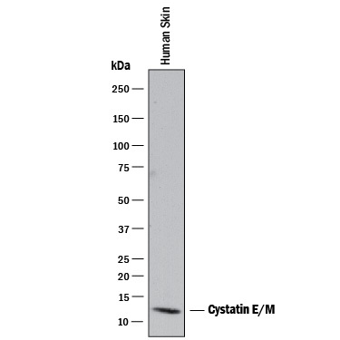
- Main image

- Experimental details
- Detection of Human Cystatin E/M by Western Blot. Western blot shows lysates of human skin tissue. PVDF membrane was probed with 1 µg/mL of Goat Anti-Human Cystatin E/M Antigen Affinity-purified Polyclonal Antibody (Catalog # AF1286) followed by HRP-conjugated Anti-Goat IgG Secondary Antibody (Catalog # HAF017). A specific band was detected for Cystatin E/M at approximately 13 kDa (as indicated). This experiment was conducted under reducing conditions and using Immunoblot Buffer Group 1.
- Submitted by
- Novus Biologicals (provider)
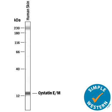
- Main image

- Experimental details
- Detection of Human Cystatin E/M by Simple WesternTM. Simple Western lane view shows lysates of human skin tissue, loaded at 0.2 mg/mL. A specific band was detected for Cystatin E/M at approximately 13 kDa (as indicated) using 50 µg/mL of Goat Anti-Human Cystatin E/M Antigen Affinity-purified Polyclonal Antibody (Catalog # AF1286) followed by 1:50 dilution of HRP-conjugated Anti-Goat IgG Secondary Antibody (Catalog # HAF109). This experiment was conducted under reducing conditions and using the 12-230 kDa separation system. Non-specific interaction with the 230 kDa Simple Western standard may be seen with this antibody.