Explore
 Validate
 Learn
Learn Immunocytochemistry
 Immunohistochemistry
Antibody data
- Antibody Data
- Antigen structure
- References [1]
- Comments [0]
- Validations
- Immunocytochemistry [1]
Submit
Validation data
Reference
Comment
Report error
- Product number
- AMAb91163 - Provider product page

- Provider
- Atlas Antibodies
- Proper citation
- Atlas Antibodies Cat#AMAb91163, RRID:AB_2665827
- Product name
- Anti-CDK5RAP2
- Antibody type
- Monoclonal
- Description
- Monoclonal Antibody against Human CDK5RAP2, Clone ID: CL3392, Gene description: CDK5 regulatory subunit associated protein 2, Alternative Gene Names: CEP215, KIAA1633, Validated applications: IHC, ICC, Uniprot ID: Q96SN8, Storage: Store at +4°C for short term storage. Long time storage is recommended at -20°C.
- Reactivity
- Human
- Host
- Mouse
- Conjugate
- Unconjugated
- Isotype
- IgG
- Antibody clone number
- CL3392
- Vial size
- 100 µl
- Concentration
- 0.5 mg/ml
- Storage
- Store at +4°C for short term storage. Long time storage is recommended at -20°C.
- Handling
- The antibody solution should be gently mixed before use.
Submitted references Pathogenic LRRK2 regulates centrosome cohesion via Rab10/RILPL1-mediated CDK5RAP2 displacement.
Fdez E, Madero-Pérez J, Lara Ordóñez AJ, Naaldijk Y, Fasiczka R, Aiastui A, Ruiz-Martínez J, López de Munain A, Cowley SA, Wade-Martins R, Hilfiker S
iScience 2022 Jun 17;25(6):104476
iScience 2022 Jun 17;25(6):104476
No comments: Submit comment
Supportive validation
- Submitted by
- Atlas Antibodies (provider)
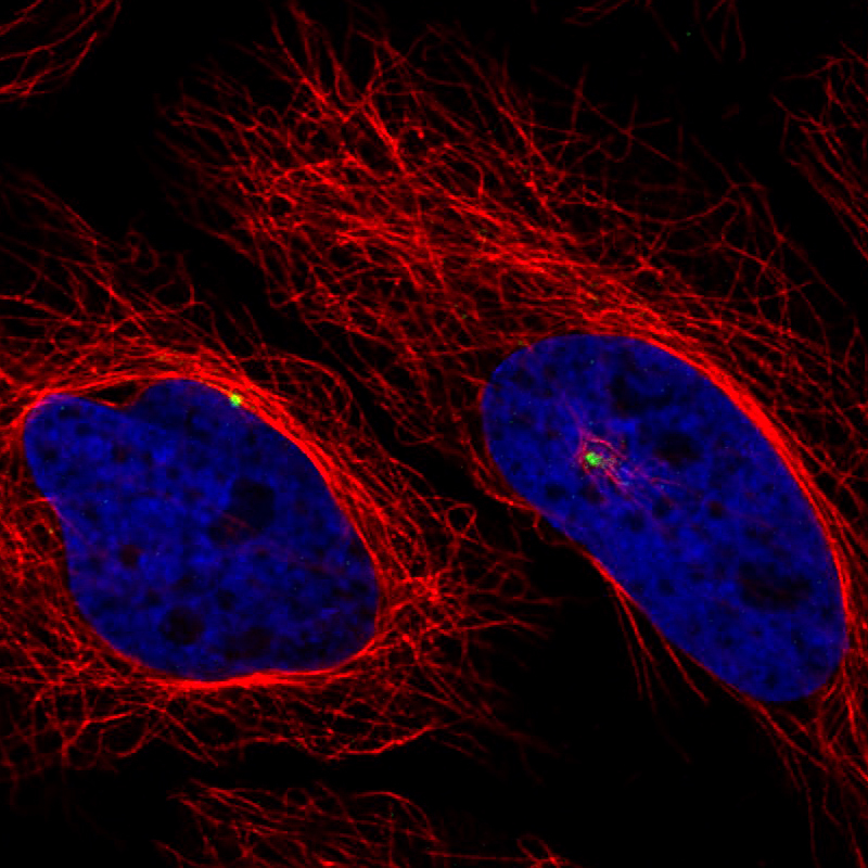
- Main image
- Experimental details
- Immunofluorescence staining of HeLa cells using the anti-CDK5RAP2 monoclonal antibody, showing specific staining of the centrosome in green. Microtubule- and nuclear probes are visualized in red and blue, respectively (where available).
- Sample type
- Human