Explore
 Validate
 Learn
Learn Western blot
Antibody data
- Antibody Data
- Antigen structure
- References [0]
- Comments [0]
- Validations
- Western blot [1]
- Immunocytochemistry [1]
- Immunohistochemistry [1]
Submit
Validation data
Reference
Comment
Report error
- Product number
- AGC-038-200UL - Provider product page

- Provider
- Invitrogen Antibodies
- Product name
- GRID1 (extracellular) Polyclonal Antibody
- Antibody type
- Polyclonal
- Antigen
- Other
- Reactivity
- Human, Mouse, Rat
- Host
- Rabbit
- Isotype
- IgG
- Vial size
- 200 µL
- Concentration
- 0.8 mg/mL
- Storage
- -20° C, Avoid Freeze/Thaw Cycles
No comments: Submit comment
Supportive validation
- Submitted by
- Invitrogen Antibodies (provider)
- Main image

- Experimental details
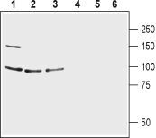
- Western blot analysis of human CCF-STTG1 astrocytoma cell lysate (lanes 1 and 4), mouse brain lysate (lanes 2 and 5) and rat brain lysate (lines 3 and 6): - 1-3. Anti-GRID1 (extracellular) Antibody (#AGC-038), (1:200).4-6. Anti-GRID1 (extracellular) Antibody , preincubated with GRID1 (extracellular) Blocking Peptide (#BLP-GC038).
Supportive validation
- Submitted by
- Invitrogen Antibodies (provider)
- Main image

- Experimental details
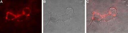
- Expression of Glutamate receptor delta 1 in rat PC12 cells - Cell surface detection of GluD1 in live intact rat PC12 pheochromocytoma cells. A. Extracellular staining of cells with Anti-GRID1 (extracellular) Antibody (#AGC-038), (1:50), followed by goat Anti-rabbit-AlexaFluor-594 secondary Antibody (red). B. Live view of the cells. C. Merge of A and B.
Supportive validation
- Submitted by
- Invitrogen Antibodies (provider)
- Main image
- Experimental details
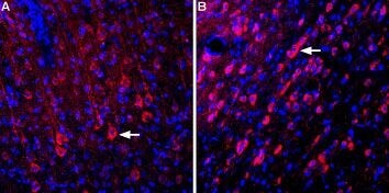
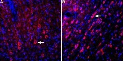
- Expression of Glutamate receptor delta 1 in rat cortex and medial septum - Immunohistochemical staining of perfusion-fixed frozen rat brain sections using Anti-GRID1 (extracellular) Antibody (#AGC-038), (1:400). A. Staining in cortex. B. Staining in medial septum. In both regions, GluD1 expression (red) is detected in neurons (arrows). DAPI is used as the counterstain (blue).