 Explore
 Validate
 Learn
Learn Western blot
 Immunocytochemistry
Antibody data
- Antibody Data
- Antigen structure
- References [1]
- Comments [0]
- Validations
- Immunocytochemistry [2]
- Immunohistochemistry [1]
Submit
Validation data
Reference
Comment
Report error
- Product number
- MA5-15537 - Provider product page

- Provider
- Invitrogen Antibodies
- Product name
- CD73 Monoclonal Antibody (1D7)
- Antibody type
- Monoclonal
- Antigen
- Purifed from natural sources
- Description
- MA5-15537 targets NT5E in IHC applications and shows reactivity with Human samples. The MA5-15537 immunogen is purified recombinant fragment of NT5E expressed in E. Coli. MA5-15537 detects NT5E which has a predicted molecular weight of approximately 70kDa.
- Reactivity
- Human
- Host
- Mouse
- Isotype
- IgG
- Antibody clone number
- 1D7
- Vial size
- 100 μL
- Concentration
- Conc. Not Determined
- Storage
- Store at 4°C short term. For long term storage, store at -20°C, avoiding freeze/thaw cycles.
Submitted references Resolvin D1 Modulates the Intracellular VEGF-Related miRNAs of Retinal Photoreceptors Challenged With High Glucose.
Maisto R, Trotta MC, Petrillo F, Izzo S, Cuomo G, Alfano R, Hermenean A, Barcia JM, Galdiero M, Platania CBM, Bucolo C, D'Amico M
Frontiers in pharmacology 2020;11:235
Frontiers in pharmacology 2020;11:235
No comments: Submit comment
Supportive validation
- Submitted by
- Invitrogen Antibodies (provider)
- Main image

- Experimental details
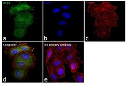
- Immunofluorescence analysis of CD73 was performed using 70% confluent log phase A-375 cells. The cells were fixed with 4% paraformaldehyde for 10 minutes, permeabilized with 0.1% Triton™ X-100 for 15 minutes, and blocked with 1% BSA for 1 hour at room temperature. The cells were labeled with CD73 Monoclonal Antibody (1D7) (Product # MA5-15537) at 1:100 dilution in 0.1% BSA, incubated at 4 degree Celsius overnight and then labeled with Goat anti-Mouse IgG (H+L) Superclonal™ Secondary Antibody, Alexa Fluor® 488 conjugate (Product # A28175) at a dilution of 1:2000 for 45 minutes at room temperature (Panel a: green). Nuclei (Panel b: blue) were stained with ProLong™ Diamond Antifade Mountant with DAPI (Product # P36962). F-actin (Panel c: red) was stained with Rhodamine Phalloidin (Product # R415). Panel d represents the merged image showing membranous and cytosolic localization. The images were captured at 60X magnification.

- Submitted by
- Invitrogen Antibodies (provider)
- Main image
- Experimental details
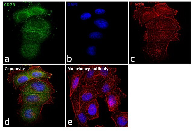
- Immunofluorescence analysis of CD73 was performed using 70% confluent log phase A-375 cells. The cells were fixed with 4% paraformaldehyde for 10 minutes, permeabilized with 0.1% Triton™ X-100 for 15 minutes, and blocked with 1% BSA for 1 hour at room temperature. The cells were labeled with CD73 Monoclonal Antibody (1D7) (Product # MA5-15537) at 1:100 dilution in 0.1% BSA, incubated at 4 degree Celsius overnight and then labeled with Goat anti-Mouse IgG (H+L) Superclonal™ Secondary Antibody, Alexa Fluor® 488 conjugate (Product # A28175) at a dilution of 1:2000 for 45 minutes at room temperature (Panel a: green). Nuclei (Panel b: blue) were stained with ProLong™ Diamond Antifade Mountant with DAPI (Product # P36962). F-actin (Panel c: red) was stained with Rhodamine Phalloidin (Product # R415). Panel d represents the merged image showing membranous and cytosolic localization. The images were captured at 60X magnification.
Supportive validation
- Submitted by
- Invitrogen Antibodies (provider)
- Main image
- Experimental details
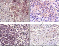
- Immunohistochemical analysis of paraffin-embedded human lung cancer (A), cholangiocarcinoma (B), lymph node (C) and esophagus (D) tissues using CD73/NT5E monoclonal antibody (Product # MA5-15537) followed with DAB staining.
