Explore
 Validate
 Learn
Learn Western blot
Antibody data
- Antibody Data
- Antigen structure
- References [2]
- Comments [0]
- Validations
- Western blot [1]
- Immunohistochemistry [1]
- Flow cytometry [1]
Submit
Validation data
Reference
Comment
Report error
- Product number
- AF5375 - Provider product page

- Provider
- R&D Systems
- Product name
- Mouse Plexin C1 Antibody
- Antibody type
- Polyclonal
- Description
- Antigen Affinity-purified. Detects mouse Plexin C1 in direct ELISAs and Western blots. In direct ELISAs, approximately 50% cross-reactivity with recombinant human Plexin C1 is observed.
- Reactivity
- Mouse
- Host
- Sheep
- Conjugate
- Unconjugated
- Antigen sequence
Q9QZC2- Isotype
- IgG
- Vial size
- 100 ug
- Concentration
- LYOPH
- Storage
- Use a manual defrost freezer and avoid repeated freeze-thaw cycles. 12 months from date of receipt, -20 to -70 °C as supplied. 1 month, 2 to 8 °C under sterile conditions after reconstitution. 6 months, -20 to -70 °C under sterile conditions after reconstitution.
Submitted references Rewiring the taste system.
Unbiased classification of sensory neuron types by large-scale single-cell RNA sequencing.
Lee H, Macpherson LJ, Parada CA, Zuker CS, Ryba NJP
Nature 2017 Aug 17;548(7667):330-333
Nature 2017 Aug 17;548(7667):330-333
Unbiased classification of sensory neuron types by large-scale single-cell RNA sequencing.
Usoskin D, Furlan A, Islam S, Abdo H, Lönnerberg P, Lou D, Hjerling-Leffler J, Haeggström J, Kharchenko O, Kharchenko PV, Linnarsson S, Ernfors P
Nature neuroscience 2015 Jan;18(1):145-53
Nature neuroscience 2015 Jan;18(1):145-53
No comments: Submit comment
Supportive validation
- Submitted by
- R&D Systems (provider)
- Main image
- Experimental details
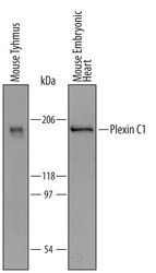
- Detection of Mouse Plexin C1 by Western Blot. Western blot shows lysates of mouse thymus tissue and embryonic mouse heart tissue. PVDF membrane was probed with 1 µg/mL of Sheep Anti-Mouse Plexin C1 Antigen Affinity-purified Polyclonal Antibody (Catalog # AF5375) followed by HRP-conjugated Anti-Sheep IgG Secondary Antibody (Catalog # HAF016). A specific band was detected for Plexin C1 at approximately 200 kDa (as indicated). This experiment was conducted under reducing conditions and using Immunoblot Buffer Group 8.
Supportive validation
- Submitted by
- R&D Systems (provider)
- Main image
- Experimental details
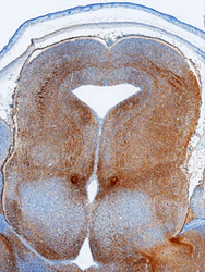
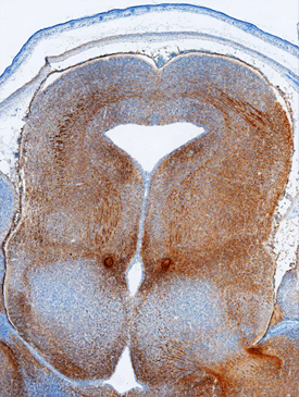
- Plexin C1 in Mouse Embryo. Plexin C1 was detected in immersion fixed frozen sections of mouse embryo (15 d.p.c.) using Sheep Anti-Mouse Plexin C1 Antigen Affinity-purified Polyclonal Antibody (Catalog # AF5375) at 5 µg/mL overnight at 4 °C. Tissue was stained using the Anti-Sheep HRP-DAB Cell & Tissue Staining Kit (brown; Catalog # CTS019) and counterstained with hematoxylin (blue). Specific staining was localized to neurites in the developing midbrain. View our protocol for Chromogenic IHC Staining of Frozen Tissue Sections.
Supportive validation
- Submitted by
- R&D Systems (provider)
- Main image
- Experimental details
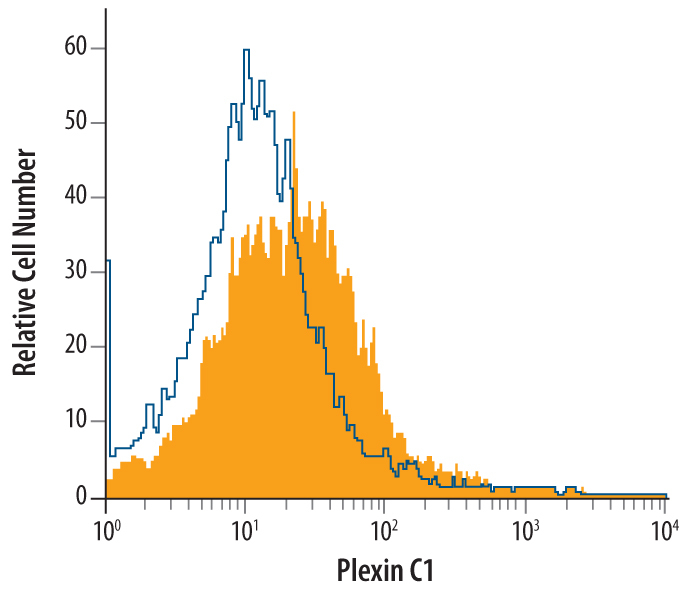
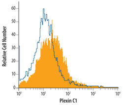
- Detection of Plexin C1 in Mouse Splenocytes by Flow Cytometry. Mouse splenocytes were stained with Sheep Anti-Mouse Plexin C1 Antigen Affinity-purified Polyclonal Antibody (Catalog # AF5375, filled histogram) or control antibody (Catalog # 5-001-A, open histogram), followed by NorthernLights™ 637-conjugated Anti-Sheep IgG Secondary Antibody (Catalog # NL011).