Explore
 Validate
 Learn
Learn Western blot
 Immunocytochemistry
Antibody data
- Antibody Data
- Antigen structure
- References [4]
- Comments [0]
- Validations
- Western blot [1]
- Immunocytochemistry [1]
Submit
Validation data
Reference
Comment
Report error
- Product number
- HPA040416 - Provider product page

- Provider
- Atlas Antibodies
- Proper citation
- Atlas Antibodies Cat#HPA040416, RRID:AB_10795524
- Product name
- Anti-KLC2
- Antibody type
- Polyclonal
- Description
- Polyclonal Antibody against Human KLC2, Gene description: kinesin light chain 2, Alternative Gene Names: FLJ12387, Validated applications: WB, ICC, Uniprot ID: Q9H0B6, Storage: Store at +4°C for short term storage. Long time storage is recommended at -20°C.
- Reactivity
- Human, Mouse, Rat
- Host
- Rabbit
- Conjugate
- Unconjugated
- Isotype
- IgG
- Vial size
- 100 µl
- Concentration
- 0.6 mg/ml
- Storage
- Store at +4°C for short term storage. Long time storage is recommended at -20°C.
- Handling
- The antibody solution should be gently mixed before use.
Submitted references Proximal protein landscapes of the type I interferon signaling cascade reveal negative regulation by PJA2.
Kinesin-1 transports morphologically distinct intracellular virions during vaccinia infection.
A Novel SLC5A5 Variant Reveals the Crucial Role of Kinesin Light Chain 2 in Thyroid Hormonogenesis.
Immunofluorescence and fluorescent-protein tagging show high correlation for protein localization in mammalian cells
Schiefer S, Hale BG
Nature communications 2024 May 27;15(1):4484
Nature communications 2024 May 27;15(1):4484
Kinesin-1 transports morphologically distinct intracellular virions during vaccinia infection.
Xu A, Basant A, Schleich S, Newsome TP, Way M
Journal of cell science 2023 Mar 1;136(5)
Journal of cell science 2023 Mar 1;136(5)
A Novel SLC5A5 Variant Reveals the Crucial Role of Kinesin Light Chain 2 in Thyroid Hormonogenesis.
Martín M, Modenutti CP, Gil Rosas ML, Peyret V, Geysels RC, Bernal Barquero CE, Sobrero G, Muñoz L, Signorino M, Testa G, Miras MB, Masini-Repiso AM, Calcaterra NB, Coux G, Carrasco N, Martí MA, Nicola JP
The Journal of clinical endocrinology and metabolism 2021 Jun 16;106(7):1867-1881
The Journal of clinical endocrinology and metabolism 2021 Jun 16;106(7):1867-1881
Immunofluorescence and fluorescent-protein tagging show high correlation for protein localization in mammalian cells
Stadler C, Rexhepaj E, Singan V, Murphy R, Pepperkok R, Uhlén M, Simpson J, Lundberg E
Nature Methods 2013;10(4):315-323
Nature Methods 2013;10(4):315-323
No comments: Submit comment
Enhanced validation
- Submitted by
- Atlas Antibodies (provider)
- Enhanced method
- Genetic validation
- Main image
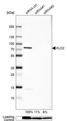
- Experimental details
- Western blot analysis in U-138MG cells transfected with control siRNA, target specific siRNA probe #1 and #2, using Anti-KLC2 antibody. Remaining relative intensity is presented. Loading control: Anti-GAPDH.
- Sample type
- Human
- Protocol
- Protocol

Supportive validation
- Submitted by
- Atlas Antibodies (provider)
- Main image

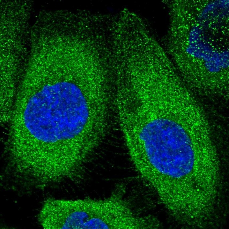
- Experimental details
- Immunofluorescent staining of human cell line A-431 shows positivity in plasma membrane & cytoplasm.
- Sample type
- Human