Explore
 Validate
 Learn
Learn Western blot
 Immunocytochemistry
Antibody data
- Antibody Data
- Antigen structure
- References [3]
- Comments [0]
- Validations
- Immunocytochemistry [8]
- Immunoprecipitation [1]
- Other assay [4]
Submit
Validation data
Reference
Comment
Report error
- Product number
- PA1-519 - Provider product page

- Provider
- Invitrogen Antibodies
- Product name
- ATRIP Polyclonal Antibody
- Antibody type
- Polyclonal
- Antigen
- Other
- Description
- PA1-519 detects human and mouse ATRIP. PA1-519 has been successfully used in Western blot, immunoprecipitation and immunofluorescence procedures. By Western blot, this antibody detects an ~ 86 kDa protein from human HeLa cells. PA1-519 antigen corresponds to amino acids 1-182 of human ATRIP
- Reactivity
- Human, Mouse
- Host
- Rabbit
- Isotype
- IgG
- Vial size
- 100 μg
- Concentration
- 1 mg/mL
- Storage
- -20°C, Avoid Freeze/Thaw Cycles
Submitted references The Atr-Chek1 pathway inhibits axon regeneration in response to Piezo-dependent mechanosensation.
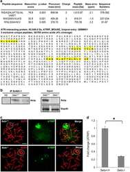
Senataxin controls meiotic silencing through ATR activation and chromatin remodeling.
Replication stress is a potent driver of functional decline in ageing haematopoietic stem cells.
Li F, Lo TY, Miles L, Wang Q, Noristani HN, Li D, Niu J, Trombley S, Goldshteyn JI, Wang C, Wang S, Qiu J, Pogoda K, Mandal K, Brewster M, Rompolas P, He Y, Janmey PA, Thomas GM, Li S, Song Y
Nature communications 2021 Jun 22;12(1):3845
Nature communications 2021 Jun 22;12(1):3845
Senataxin controls meiotic silencing through ATR activation and chromatin remodeling.
Yeo AJ, Becherel OJ, Luff JE, Graham ME, Richard D, Lavin MF
Cell discovery 2015;1:15025
Cell discovery 2015;1:15025
Replication stress is a potent driver of functional decline in ageing haematopoietic stem cells.
Flach J, Bakker ST, Mohrin M, Conroy PC, Pietras EM, Reynaud D, Alvarez S, Diolaiti ME, Ugarte F, Forsberg EC, Le Beau MM, Stohr BA, Méndez J, Morrison CG, Passegué E
Nature 2014 Aug 14;512(7513):198-202
Nature 2014 Aug 14;512(7513):198-202
No comments: Submit comment
Supportive validation
- Submitted by
- Invitrogen Antibodies (provider)
- Main image

- Experimental details
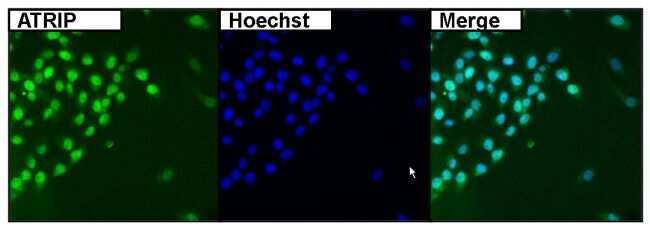
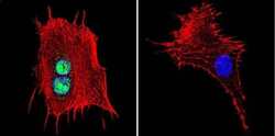
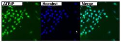
- Immunofluorescent analysis of ATRIP (green) in HeLa cells. Formalin fixed cells were permeabilized with 0.1% Triton X-100 in TBS for 10 minutes at room temperature. Cells were then blocked with 5% normal goat serum (Product # 31873) for 15 minutes at room temperature. Cells were then probed with a rabbit polyclonal antibody recognizing ATRIP (Product # PA1-519), at a dilution of 1:400 for at least 1 hour at room temperature. Cells were then washed with PBS and incubated with DyLight 488 goat-anti-rabbit secondary antibody at a dilution of 1:400 for 30 minutes at room temperature. Nuclei (blue) were stained with Hoechst 33342 dye (Product # 62249). Images were taken on a Thermo Scientific ArrayScan at 20X magnification.
- Submitted by
- Invitrogen Antibodies (provider)
- Main image
- Experimental details
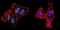
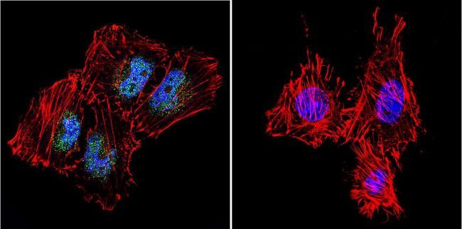
- Immunofluorescent analysis of ATRIP in HeLa Cells. Cells were grown on chamber slides and fixed with formaldehyde prior to staining. Cells were probed without (control) or with a ATRIP polyclonal antibody (Product # PA1-519) at a dilution of 1:100 overnight at 4 C, washed with PBS and incubated with a DyLight-488 conjugated secondary antibody (Product # 35552). ATRIP staining (green), F-Actin staining with Phalloidin (red) and nuclei with DAPI (blue) is shown. Images were taken at 60X magnification.

- Submitted by
- Invitrogen Antibodies (provider)
- Main image

- Experimental details
- Immunofluorescent analysis of ATRIP in NIH-3T3 Cells. Cells were grown on chamber slides and fixed with formaldehyde prior to staining. Cells were probed without (control) or with a ATRIP polyclonal antibody (Product # PA1-519) at a dilution of 1:100 overnight at 4 C, washed with PBS and incubated with a DyLight-488 conjugated secondary antibody (Product # 35552). ATRIP staining (green), F-Actin staining with Phalloidin (red) and nuclei with DAPI (blue) is shown. Images were taken at 60X magnification.

- Submitted by
- Invitrogen Antibodies (provider)
- Main image

- Experimental details
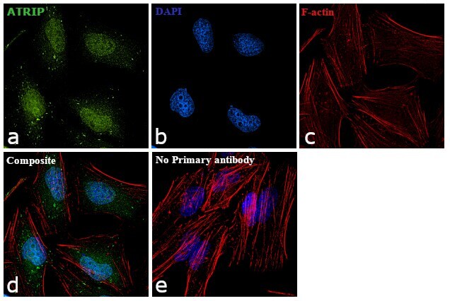
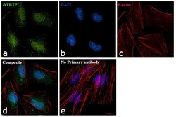
- Immunofluorescence analysis of ATRIP was performed using 70% confluent log phase HeLa cells. The cells were fixed with 4% paraformaldehyde for 10 minutes, permeabilized with 0.1% Triton™ X-100 for 15 minutes, and blocked with 1% BSA for 1 hour at room temperature. The cells were labeled with ATRIP Polyclonal Antibody (Product # PA1-519) at 5µg/mL in 0.1% BSA, incubated at 4 degree Celsius overnight and then labeled with Goat anti-Rabbit IgG (H+L) Superclonal™ Secondary Antibody, Alexa Fluor® 488 conjugate (Product # A27034) at a dilution of 1:2000 for 45 minutes at room temperature (Panel a: green). Nuclei (Panel b: blue) were stained with ProLong™ Diamond Antifade Mountant with DAPI (Product # P36962). F-actin (Panel c: red) was stained with Rhodamine Phalloidin (Product # R415). Panel d represents the merged image showing Nuclear localization. Panel e represents control cells with no primary antibody to assess background. The images were captured at 60X magnification.
- Submitted by
- Invitrogen Antibodies (provider)
- Main image

- Experimental details
- Immunofluorescent analysis of ATRIP in HeLa Cells. Cells were grown on chamber slides and fixed with formaldehyde prior to staining. Cells were probed without (control) or with a ATRIP polyclonal antibody (Product # PA1-519) at a dilution of 1:100 overnight at 4 C, washed with PBS and incubated with a DyLight-488 conjugated secondary antibody (Product # 35552). ATRIP staining (green), F-Actin staining with Phalloidin (red) and nuclei with DAPI (blue) is shown. Images were taken at 60X magnification.
- Submitted by
- Invitrogen Antibodies (provider)
- Main image
- Experimental details
- Immunofluorescent analysis of ATRIP in NIH-3T3 Cells. Cells were grown on chamber slides and fixed with formaldehyde prior to staining. Cells were probed without (control) or with a ATRIP polyclonal antibody (Product # PA1-519) at a dilution of 1:100 overnight at 4 C, washed with PBS and incubated with a DyLight-488 conjugated secondary antibody (Product # 35552). ATRIP staining (green), F-Actin staining with Phalloidin (red) and nuclei with DAPI (blue) is shown. Images were taken at 60X magnification.

- Submitted by
- Invitrogen Antibodies (provider)
- Main image
- Experimental details
- Immunofluorescent analysis of ATRIP (green) in HeLa cells. Formalin fixed cells were permeabilized with 0.1% Triton X-100 in TBS for 10 minutes at room temperature. Cells were then blocked with 5% normal goat serum (Product # 31873) for 15 minutes at room temperature. Cells were then probed with a rabbit polyclonal antibody recognizing ATRIP (Product # PA1-519), at a dilution of 1:400 for at least 1 hour at room temperature. Cells were then washed with PBS and incubated with DyLight 488 goat-anti-rabbit secondary antibody at a dilution of 1:400 for 30 minutes at room temperature. Nuclei (blue) were stained with Hoechst 33342 dye (Product # 62249). Images were taken on a Thermo Scientific ArrayScan at 20X magnification.

- Submitted by
- Invitrogen Antibodies (provider)
- Main image
- Experimental details
- Immunofluorescence analysis of ATRIP was performed using 70% confluent log phase HeLa cells. The cells were fixed with 4% paraformaldehyde for 10 minutes, permeabilized with 0.1% Triton™ X-100 for 15 minutes, and blocked with 1% BSA for 1 hour at room temperature. The cells were labeled with ATRIP Polyclonal Antibody (Product # PA1-519) at 5µg/mL in 0.1% BSA, incubated at 4 degree Celsius overnight and then labeled with Goat anti-Rabbit IgG (Heavy Chain) Superclonal™ Secondary Antibody, Alexa Fluor® 488 conjugate (Product # A27034) at a dilution of 1:2000 for 45 minutes at room temperature (Panel a: green). Nuclei (Panel b: blue) were stained with ProLong™ Diamond Antifade Mountant with DAPI (Product # P36962). F-actin (Panel c: red) was stained with Rhodamine Phalloidin (Product # R415). Panel d represents the merged image showing Nuclear localization. Panel e represents control cells with no primary antibody to assess background. The images were captured at 60X magnification.

Supportive validation
- Submitted by
- Invitrogen Antibodies (provider)
- Main image
- Experimental details
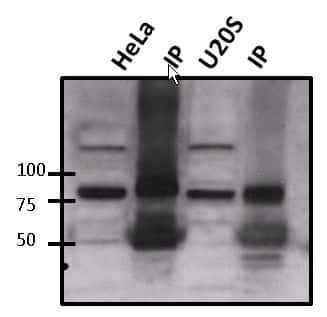
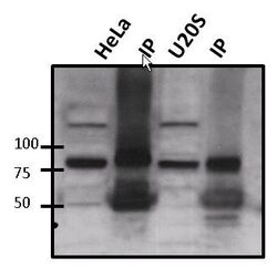
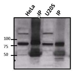
- Immunoprecipitation of ATRIP was performed on HeLa and U20S lysate. The antigen:antbody complex was formed by binding 500 µg whole cell lysate with 2 µg of rabbit polyclonal antibody recognizing ATRIP (Product # PA1-519) overnight on a rocking platform at 4°C. The immune-complex was then captured on 50 µL Protein A/G Plus Agarose (Product # 20423). Captured immune-complexes were then washed extensively and proteins eluted with 5X Reducing Sample Loading Dye (Product # 39000). Samples were then resolved on a 4-20% Tris-HCl polyacrylamide gel. Proteins were transferred to PVDF membrane and blocked with 5% Milk/TBS-0.1%Tween for at least 1 hour. Membranes were then probed with a rabbit polyclonal antibody recognizing ATRIP (Product # PA1-519) at a dilution of 1:1000 overnight rotating at 4°C. Membranes were then washed in TBST and probed with a goat anti-rabbit-HRP secondary antibody (Product # 31460) at a dilution of 1:20000 for at least one hour. Membranes were washed and chemiluminescent detection was performed using Pierce Super Signal West Pico (Product # 34077).
Supportive validation
- Submitted by
- Invitrogen Antibodies (provider)
- Main image
- Experimental details
- NULL
- Submitted by
- Invitrogen Antibodies (provider)
- Main image
- Experimental details
- NULL
- Submitted by
- Invitrogen Antibodies (provider)
- Main image
- Experimental details
- NULL

- Submitted by
- Invitrogen Antibodies (provider)
- Main image
- Experimental details
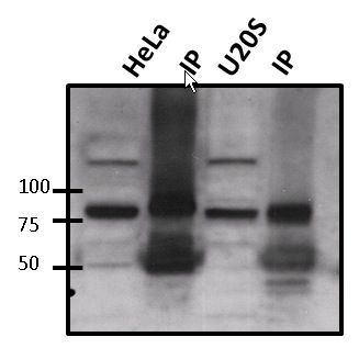
- Immunoprecipitation of ATRIP was performed on HeLa and U20S lysate. The antigen:antbody complex was formed by binding 500 µg whole cell lysate with 2 µg of rabbit polyclonal antibody recognizing ATRIP (Product # PA1-519) overnight on a rocking platform at 4øC. The immune-complex was then captured on 50 µL Protein A/G Plus Agarose (Product # 20423). Captured immune-complexes were then washed extensively and proteins eluted with 5X Reducing Sample Loading Dye (Product # 39000). Samples were then resolved on a 4-20% Tris-HCl polyacrylamide gel. Proteins were transferred to PVDF membrane and blocked with 5% Milk/TBS-0.1%Tween for at least 1 hour. Membranes were then probed with a rabbit polyclonal antibody recognizing ATRIP (Product # PA1-519) at a dilution of 1:1000 overnight rotating at 4øC. Membranes were then washed in TBST and probed with a goat anti-rabbit-HRP secondary antibody (Product # 31460) at a dilution of 1:20000 for at least one hour. Membranes were washed and chemiluminescent detection was performed using Pierce Super Signal West Pico (Product # 34077).