Explore
 Validate
 Learn
LearnNBP1-28900
antibody from Novus Biologicals
Targeting: PEG10
HB-1, KIAA1051, Mar2, Mart2, MEF3L, RGAG3, RTL2, SIRH1
 Western blot
 ELISA
ELISAAntibody data
- Antibody Data
- Antigen structure
- References [1]
- Comments [0]
- Validations
- Western blot [1]
- Immunohistochemistry [2]
Submit
Validation data
Reference
Comment
Report error
- Product number
- NBP1-28900 - Provider product page

- Provider
- Novus Biologicals
- Proper citation
- Novus Cat#NBP1-28900, RRID:AB_1914053
- Product name
- Mouse Monoclonal PEG10 Antibody
- Antibody type
- Monoclonal
- Description
- Unpurified.
- Reactivity
- Human, Mouse
- Host
- Mouse
- Isotype
- IgG
- Vial size
- 0.1 ml
- Storage
- Store at 4C short term. Aliquot and store at -20C long term. Avoid freeze-thaw cycles.
Submitted references DREAM and RB cooperate to induce gene repression and cell-cycle arrest in response to p53 activation.
Uxa S, Bernhart SH, Mages CFS, Fischer M, Kohler R, Hoffmann S, Stadler PF, Engeland K, Müller GA
Nucleic acids research 2019 Sep 26;47(17):9087-9103
Nucleic acids research 2019 Sep 26;47(17):9087-9103
No comments: Submit comment
Supportive validation
- Submitted by
- Novus Biologicals (provider)
- Main image
- Experimental details
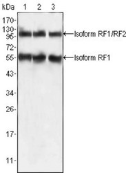
- Western Blot: PEG10 Antibody (4C10A7) [NBP1-28900] - Western blot analysis using PEG10 mouse mAb against HepG2 (1), SMMC-7721 (2) and A549 (3) cell lysate.
Supportive validation
- Submitted by
- Novus Biologicals (provider)
- Main image
- Experimental details
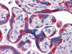
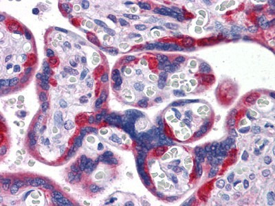
- Immunohistochemistry-Paraffin: PEG10 Antibody (4C10A7) [NBP1-28900] - Immunohistochemical analysis of paraffin-embedded human Placenta tissues using PEG10 mouse mAb
- Submitted by
- Novus Biologicals (provider)
- Main image
- Experimental details
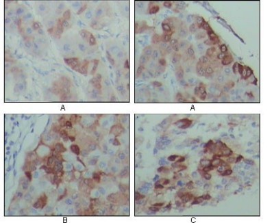
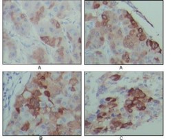
- Immunohistochemistry-Paraffin: PEG10 Antibody (4C10A7) [NBP1-28900] - Immunohistochemical analysis of paraffin-embedded human hepatocarcinoma (A), breast carcinoma (B) and lung cancer tissues (C), showing cytoplasmic localization with DAB staining using PEG10 mouse mAb.