Explore
 Validate
 Learn
LearnPA5-26365
antibody from Invitrogen Antibodies
Targeting: TRERF1
BCAR2, dJ139D8.5, HSA277276, RAPA, TReP-132
 Western blot
 Immunocytochemistry
Antibody data
- Antibody Data
- Antigen structure
- References [0]
- Comments [0]
- Validations
- Immunocytochemistry [2]
- Flow cytometry [2]
Submit
Validation data
Reference
Comment
Report error
- Product number
- PA5-26365 - Provider product page

- Provider
- Invitrogen Antibodies
- Product name
- TREF1 Polyclonal Antibody
- Antibody type
- Polyclonal
- Antigen
- Synthetic peptide
- Reactivity
- Human
- Host
- Rabbit
- Isotype
- IgG
- Vial size
- 400 μL
- Storage
- Store at 4°C short term. For long term storage, store at -20°C, avoiding freeze/thaw cycles.
No comments: Submit comment
Supportive validation
- Submitted by
- Invitrogen Antibodies (provider)
- Main image

- Experimental details
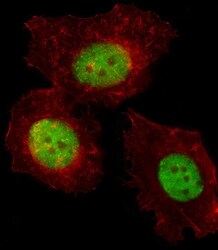
- Immunofluorescent analysis of MCF-7 cells using a TREF1 polyclonal antibody (Product # PA5-26365). MCF-7 cells were fixed with 4% PFA (20 min), permeabilized with Triton X-100 (0.1%, 10 min), then incubated with a TREF1 polyclonal antibody (Product # PA5-26365) (1:25, 1 hr at 37ºC). Primary antibody was detected with fluor-conjugated donkey anti-rabbit secondary antibody (green) at 1:400 dilution for 50 min at 37ºC). Actin filaments have been labeled with dye-conjugated phalloidin (red).

- Submitted by
- Invitrogen Antibodies (provider)
- Main image
- Experimental details
- Immunocytochemistry analysis of TREF1 in MCF-7 cells. Samples were incubated with TREF1 polyclonal antibody (Product # PA5-26365) using a dilution of 1:25 for 1 h at 37°C followed by Alexa Fluor® 488 conjugated donkey anti-rabbit antibody (green) at a dilution of 1:400 for 50 min at 37°C. Cells were fixed with 4% PFA (20 min) and permeabilized with Triton X-100 (0.1%, 10 min). Cytoplasmic actin was counterstained with Alexa Fluor® 555 (red) conjugated Phalloidin (7 units/mL, 1 h at 37°C). TREF1 immunoreactivity is localized to Nucleus significantly.

Supportive validation
- Submitted by
- Invitrogen Antibodies (provider)
- Main image
- Experimental details
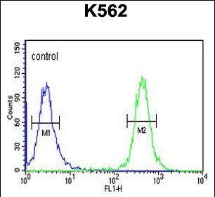
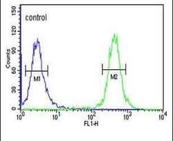
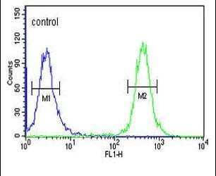
- Flow cytometry analysis of K562 cells using a TREF1 polyclonal antibody (Product # PA5-26365) (right) compared to a negative control cell (left) at a dilution of 1:10-50, followed by a FITC-conjugated goat anti-rabbit antibody
- Submitted by
- Invitrogen Antibodies (provider)
- Main image
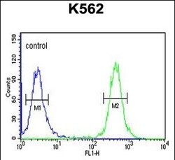
- Experimental details
- Flow cytometry of TREF1 in K562 cells (right histogram). Samples were incubated with TREF1 polyclonal antibody (Product # PA5-26365) followed by FITC-conjugated goat-anti-rabbit secondary antibody. Negative control cell (left histogram).