Explore
 Validate
 Learn
Learn13639-1-AP
antibody from Proteintech Group
Targeting: SEM1
C7orf76, DSS1, ECD, FLJ42280, SHFD1, Shfdg1, SHFM1, SHSF1
 Western blot
 ELISA
ELISAAntibody data
- Antibody Data
- Antigen structure
- References [11]
- Comments [0]
- Validations
- Western blot [1]
- Immunocytochemistry [1]
Submit
Validation data
Reference
Comment
Report error
- Product number
- 13639-1-AP - Provider product page

- Provider
- Proteintech Group
- Proper citation
- Proteintech Cat#13639-1-AP, RRID:AB_2254633
- Product name
- DSS1 antibody
- Antibody type
- Polyclonal
- Description
- KD/KO validated DSS1 antibody (Cat. #13639-1-AP) is a rabbit polyclonal antibody that shows reactivity with human and has been validated for the following applications: IHC,ELISA.
- Reactivity
- Human
- Host
- Rabbit
- Conjugate
- Unconjugated
- Isotype
- IgG
- Vial size
- 20ul, 150ul
Submitted references DSS1 is required for proper Integrator-PP2A function.
DSS1 inhibits autophagy to activate epithelial-mesenchymal transition in a pro-metastatic niche of renal cell carcinoma.
SHFM1 deficiency suppresses esophageal squamous cell carcinomas progression via modulating NF‑κB signaling and enhancing nature killer cell‑mediated tumor surveillance.
Evidence for reduced BRCA2 functional activity in Homo sapiens after divergence from the chimpanzee-human last common ancestor.
Increased chemosensitivity via BRCA2-independent DNA damage in DSS1- and PCID2-depleted breast carcinomas.
DSS1 promoter hypomethylation and overexpression predict poor prognosis in melanoma and squamous cell carcinoma patients.
BRCA2 prevents R-loop accumulation and associates with TREX-2 mRNA export factor PCID2.
DSSylation, a novel protein modification targets proteins induced by oxidative stress, and facilitates their degradation in cells.
Absent expression of the osteoblast-specific maternally imprinted genes, DLX5 and DLX6, causes split hand/split foot malformation type I.
Breast cancers with high DSS1 expression that potentially maintains BRCA2 stability have poor prognosis in the relapse-free survival.
The proteasomal de-ubiquitinating enzyme POH1 promotes the double-strand DNA break response.
Xu C, Zhou QX, Zheng H, Song A, Zhao WY, Xu TT, Xiong Y, Zhang YJ, Huang Z, Xu Y, Cheng J, Chen FX
Nature communications 2025 Jul 5;16(1):6206
Nature communications 2025 Jul 5;16(1):6206
DSS1 inhibits autophagy to activate epithelial-mesenchymal transition in a pro-metastatic niche of renal cell carcinoma.
Chen X, Liu Q, Wu J, Zhou P, Zhao M, Song J
Nature communications 2025 Jul 23;16(1):6769
Nature communications 2025 Jul 23;16(1):6769
SHFM1 deficiency suppresses esophageal squamous cell carcinomas progression via modulating NF‑κB signaling and enhancing nature killer cell‑mediated tumor surveillance.
Wu Y, Wang Z, Li S, Chen X, Zhou S
Experimental and therapeutic medicine 2023 May;25(5):195
Experimental and therapeutic medicine 2023 May;25(5):195
Evidence for reduced BRCA2 functional activity in Homo sapiens after divergence from the chimpanzee-human last common ancestor.
Huang J, Zhong Y, Makohon-Moore AP, White T, Jasin M, Norell MA, Wheeler WC, Iacobuzio-Donahue CA
Cell reports 2022 May 3;39(5):110771
Cell reports 2022 May 3;39(5):110771
Increased chemosensitivity via BRCA2-independent DNA damage in DSS1- and PCID2-depleted breast carcinomas.
Gondo N, Sakai Y, Zhang Z, Hato Y, Kuzushima K, Phimsen S, Kawashima Y, Kuroda M, Suzuki M, Okada S, Iwata H, Toyama T, Rezano A, Kuwahara K
Laboratory investigation; a journal of technical methods and pathology 2021 Aug;101(8):1048-1059
Laboratory investigation; a journal of technical methods and pathology 2021 Aug;101(8):1048-1059
DSS1 promoter hypomethylation and overexpression predict poor prognosis in melanoma and squamous cell carcinoma patients.
Venza M, Visalli M, Catalano T, Beninati C, Teti D, Venza I
Human pathology 2017 Feb;60:137-146
Human pathology 2017 Feb;60:137-146
BRCA2 prevents R-loop accumulation and associates with TREX-2 mRNA export factor PCID2.
Bhatia V, Barroso SI, García-Rubio ML, Tumini E, Herrera-Moyano E, Aguilera A
Nature 2014 Jul 17;511(7509):362-5
Nature 2014 Jul 17;511(7509):362-5
DSSylation, a novel protein modification targets proteins induced by oxidative stress, and facilitates their degradation in cells.
Zhang Y, Chang FM, Huang J, Junco JJ, Maffi SK, Pridgen HI, Catano G, Dang H, Ding X, Yang F, Kim DJ, Slaga TJ, He R, Wei SJ
Protein & cell 2014 Feb;5(2):124-40
Protein & cell 2014 Feb;5(2):124-40
Absent expression of the osteoblast-specific maternally imprinted genes, DLX5 and DLX6, causes split hand/split foot malformation type I.
Rattanasopha S, Tongkobpetch S, Srichomthong C, Kitidumrongsook P, Suphapeetiporn K, Shotelersuk V
Journal of medical genetics 2014 Dec;51(12):817-23
Journal of medical genetics 2014 Dec;51(12):817-23
Breast cancers with high DSS1 expression that potentially maintains BRCA2 stability have poor prognosis in the relapse-free survival.
Rezano A, Kuwahara K, Yamamoto-Ibusuki M, Kitabatake M, Moolthiya P, Phimsen S, Suda T, Tone S, Yamamoto Y, Iwase H, Sakaguchi N
BMC cancer 2013 Dec 1;13:562
BMC cancer 2013 Dec 1;13:562
The proteasomal de-ubiquitinating enzyme POH1 promotes the double-strand DNA break response.
Butler LR, Densham RM, Jia J, Garvin AJ, Stone HR, Shah V, Weekes D, Festy F, Beesley J, Morris JR
The EMBO journal 2012 Oct 3;31(19):3918-34
The EMBO journal 2012 Oct 3;31(19):3918-34
No comments: Submit comment
Supportive validation
- Submitted by
- Proteintech Group (provider)
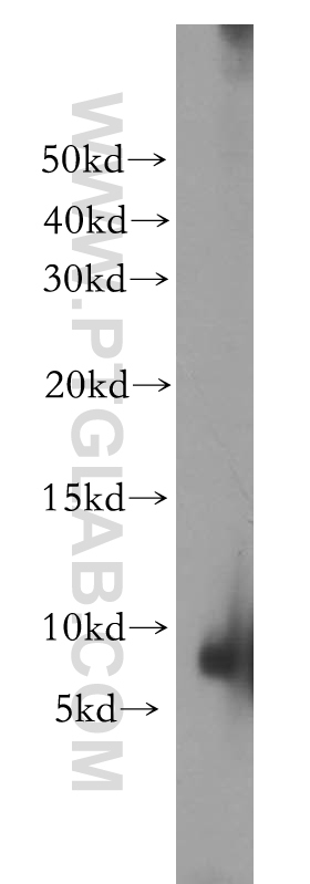
- Main image

- Experimental details
- HeLa cells were subjected to SDS PAGE followed by western blot with 13639-1-AP(SHFM1 antibody) at dilution of 1:300
- Sample type
- cell line
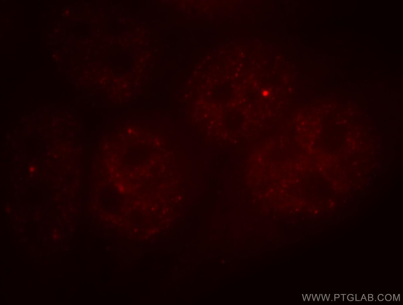
Supportive validation
- Submitted by
- Proteintech Group (provider)
- Main image
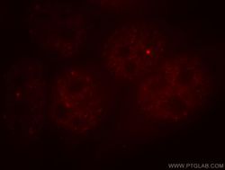
- Experimental details
- Immunofluorescent analysis of Hela cells, using SHFM1 antibody 13639-1-AP at 1:25 dilution and Rhodamine-labeled goat anti-rabbit IgG (red).
- Sample type
- cell line