Explore
 Validate
 Learn
Learn Western blot
Antibody data
- Antibody Data
- Antigen structure
- References [24]
- Comments [0]
- Validations
- Western blot [2]
- Immunocytochemistry [1]
- Immunohistochemistry [1]
- Flow cytometry [1]
- Other assay [2]
Submit
Validation data
Reference
Comment
Report error
- Product number
- PA5-16697 - Provider product page

- Provider
- Invitrogen Antibodies
- Product name
- Alpha-Smooth Muscle Actin Polyclonal Antibody
- Antibody type
- Polyclonal
- Antigen
- Synthetic peptide
- Description
- PA5-16697 targets Actin Smooth Muscle in IHC (P) applications and shows reactivity with Bovine, Chicken, Human, mouse, Non-human primate, Rabbit, and Rat samples.
- Concentration
- 0.25 mg/mL
Submitted references Data-Driven Kidney Transplant Phenotyping as a Histology-Independent Framework for Biomarker Discovery.
Protective mechanism of SIRT1 on Hcy-induced atrial fibrosis mediated by TRPC3.
Diminished Vitamin D Receptor Protein Levels in Crohn's Disease Fibroblasts: Effects of Vitamin D.
ITGA5 inhibition in pancreatic stellate cells attenuates desmoplasia and potentiates efficacy of chemotherapy in pancreatic cancer.
Global Gene Expression Analysis in an in vitro Fibroblast Model of Idiopathic Pulmonary Fibrosis Reveals Potential Role for CXCL14/CXCR4.
Prognostic Impact of Urokinase Plasminogen Activator Receptor Expression in Pancreatic Cancer: Malignant Versus Stromal Cells.
Human liver regeneration in advanced cirrhosis is organized by the portal tree.
Vascular Smooth Muscle Cells Stimulate Platelets and Facilitate Thrombus Formation through Platelet CLEC-2: Implications in Atherothrombosis.
Smooth Muscle-Targeted Overexpression of Peroxisome Proliferator Activated Receptor-γ Disrupts Vascular Wall Structure and Function.
PIK3CA(H1047R) induces multipotency and multi-lineage mammary tumours.
Compartmental and temporal dynamics of chronic inflammation and airway remodelling in a chronic asthma mouse model.
Neutralizing murine TGFβR2 promotes a differentiated tumor cell phenotype and inhibits pancreatic cancer metastasis.
Solitary tumours associated with Jaagsiekte retrovirus in sheep are heterogeneous and contain cells expressing markers identifying progenitor cells in lung repair.
The impact of KRAS mutations on VEGF-A production and tumour vascular network.
Smooth muscle cell transplantation improves bladder contractile function in streptozocin-induced diabetic rats.
The cAMP effector EPAC activates Elk1 transcription factor in prostate smooth muscle, and is a minor regulator of α1-adrenergic contraction.
Pericyte coverage of differentiated vessels inside tumor vasculature is an independent unfavorable prognostic factor for patients with clear cell renal cell carcinoma.
Generation and hepatic differentiation of human iPS cells.
Adenosine A(2a) receptor stimulation prevents hepatocyte lipotoxicity and non-alcoholic steatohepatitis (NASH) in rats.
Recruitment of podoplanin positive cancer-associated fibroblasts in metastatic lymph nodes predicts poor prognosis in pathological N2 stage III lung adenocarcinoma.
Giant cell tumor of bonelike lesion in a Trp53 mutant mouse.
Expression of NOS and VEGF in feline mammary tumours and their correlation with angiogenesis.
Deleted in colorectal carcinoma suppresses metastasis in p53-deficient mammary tumours.
Luminal expression of PIK3CA mutant H1047R in the mammary gland induces heterogeneous tumors.
Buscher K, Heitplatz B, van Marck V, Song J, Loismann S, Rixen R, Hüchtmann B, Kurian S, Ehinger E, Wolf D, Ley K, Pavenstädt H, Reuter S
Journal of the American Society of Nephrology : JASN 2021 Aug;32(8):1933-1945
Journal of the American Society of Nephrology : JASN 2021 Aug;32(8):1933-1945
Protective mechanism of SIRT1 on Hcy-induced atrial fibrosis mediated by TRPC3.
Han L, Tang Y, Li S, Wu Y, Chen X, Wu Q, Hong K, Li J
Journal of cellular and molecular medicine 2020 Jan;24(1):488-510
Journal of cellular and molecular medicine 2020 Jan;24(1):488-510
Diminished Vitamin D Receptor Protein Levels in Crohn's Disease Fibroblasts: Effects of Vitamin D.
Gisbert-Ferrándiz L, Cosín-Roger J, Hernández C, Macias-Ceja DC, Ortiz-Masiá D, Salvador P, Esplugues JV, Hinojosa J, Navarro F, Calatayud S, Barrachina MD
Nutrients 2020 Apr 1;12(4)
Nutrients 2020 Apr 1;12(4)
ITGA5 inhibition in pancreatic stellate cells attenuates desmoplasia and potentiates efficacy of chemotherapy in pancreatic cancer.
Kuninty PR, Bansal R, De Geus SWL, Mardhian DF, Schnittert J, van Baarlen J, Storm G, Bijlsma MF, van Laarhoven HW, Metselaar JM, Kuppen PJK, Vahrmeijer AL, Östman A, Sier CFM, Prakash J
Science advances 2019 Sep;5(9):eaax2770
Science advances 2019 Sep;5(9):eaax2770
Global Gene Expression Analysis in an in vitro Fibroblast Model of Idiopathic Pulmonary Fibrosis Reveals Potential Role for CXCL14/CXCR4.
Rodriguez LR, Emblom-Callahan M, Chhina M, Bui S, Aljeburry B, Tran LH, Novak R, Lemma M, Nathan SD, Grant GM
Scientific reports 2018 Mar 5;8(1):3983
Scientific reports 2018 Mar 5;8(1):3983
Prognostic Impact of Urokinase Plasminogen Activator Receptor Expression in Pancreatic Cancer: Malignant Versus Stromal Cells.
de Geus SW, Baart VM, Boonstra MC, Kuppen PJ, Prevoo HA, Mazar AP, Bonsing BA, Morreau H, van de Velde CJ, Vahrmeijer AL, Sier CF
Biomarker insights 2017;12:1177271917715443
Biomarker insights 2017;12:1177271917715443
Human liver regeneration in advanced cirrhosis is organized by the portal tree.
Dezső K, Rókusz A, Bugyik E, Szücs A, Szuák A, Dorogi B, Kiss M, Nemeskéri Á, Nagy P, Paku S
Journal of hepatology 2017 Apr;66(4):778-786
Journal of hepatology 2017 Apr;66(4):778-786
Vascular Smooth Muscle Cells Stimulate Platelets and Facilitate Thrombus Formation through Platelet CLEC-2: Implications in Atherothrombosis.
Inoue O, Hokamura K, Shirai T, Osada M, Tsukiji N, Hatakeyama K, Umemura K, Asada Y, Suzuki-Inoue K, Ozaki Y
PloS one 2015;10(9):e0139357
PloS one 2015;10(9):e0139357
Smooth Muscle-Targeted Overexpression of Peroxisome Proliferator Activated Receptor-γ Disrupts Vascular Wall Structure and Function.
Kleinhenz JM, Murphy TC, Pokutta-Paskaleva AP, Gleason RL, Lyle AN, Taylor WR, Blount MA, Cheng J, Yang Q, Sutliff RL, Hart CM
PloS one 2015;10(10):e0139756
PloS one 2015;10(10):e0139756
PIK3CA(H1047R) induces multipotency and multi-lineage mammary tumours.
Koren S, Reavie L, Couto JP, De Silva D, Stadler MB, Roloff T, Britschgi A, Eichlisberger T, Kohler H, Aina O, Cardiff RD, Bentires-Alj M
Nature 2015 Sep 3;525(7567):114-8
Nature 2015 Sep 3;525(7567):114-8
Compartmental and temporal dynamics of chronic inflammation and airway remodelling in a chronic asthma mouse model.
Alrifai M, Marsh LM, Dicke T, Kılıç A, Conrad ML, Renz H, Garn H
PloS one 2014;9(1):e85839
PloS one 2014;9(1):e85839
Neutralizing murine TGFβR2 promotes a differentiated tumor cell phenotype and inhibits pancreatic cancer metastasis.
Ostapoff KT, Cenik BK, Wang M, Ye R, Xu X, Nugent D, Hagopian MM, Topalovski M, Rivera LB, Carroll KD, Brekken RA
Cancer research 2014 Sep 15;74(18):4996-5007
Cancer research 2014 Sep 15;74(18):4996-5007
Solitary tumours associated with Jaagsiekte retrovirus in sheep are heterogeneous and contain cells expressing markers identifying progenitor cells in lung repair.
De las Heras M, de Martino A, Borobia M, Ortín A, Álvarez R, Borderías L, Giménez-Más JA
Journal of comparative pathology 2014 Feb-Apr;150(2-3):138-47
Journal of comparative pathology 2014 Feb-Apr;150(2-3):138-47
The impact of KRAS mutations on VEGF-A production and tumour vascular network.
Figueras A, Arbos MA, Quiles MT, Viñals F, Germà JR, Capellà G
BMC cancer 2013 Mar 18;13:125
BMC cancer 2013 Mar 18;13:125
Smooth muscle cell transplantation improves bladder contractile function in streptozocin-induced diabetic rats.
Gopinath C, Ponsaerts P, Fransen E, Boeykens N, Pauwels P, Wyndaele JJ
Cytotherapy 2013 Jul;15(7):869-78
Cytotherapy 2013 Jul;15(7):869-78
The cAMP effector EPAC activates Elk1 transcription factor in prostate smooth muscle, and is a minor regulator of α1-adrenergic contraction.
Hennenberg M, Strittmatter F, Schmetkamp H, Rutz B, Walther S, Stief CG, Gratzke C
Journal of biomedical science 2013 Jul 2;20(1):46
Journal of biomedical science 2013 Jul 2;20(1):46
Pericyte coverage of differentiated vessels inside tumor vasculature is an independent unfavorable prognostic factor for patients with clear cell renal cell carcinoma.
Cao Y, Zhang ZL, Zhou M, Elson P, Rini B, Aydin H, Feenstra K, Tan MH, Berghuis B, Tabbey R, Resau JH, Zhou FJ, Teh BT, Qian CN
Cancer 2013 Jan 15;119(2):313-24
Cancer 2013 Jan 15;119(2):313-24
Generation and hepatic differentiation of human iPS cells.
Ishikawa T, Hagiwara K, Ochiya T
Methods in molecular biology (Clifton, N.J.) 2012;826:103-14
Methods in molecular biology (Clifton, N.J.) 2012;826:103-14
Adenosine A(2a) receptor stimulation prevents hepatocyte lipotoxicity and non-alcoholic steatohepatitis (NASH) in rats.
Imarisio C, Alchera E, Sutti S, Valente G, Boccafoschi F, Albano E, Carini R
Clinical science (London, England : 1979) 2012 Sep;123(5):323-32
Clinical science (London, England : 1979) 2012 Sep;123(5):323-32
Recruitment of podoplanin positive cancer-associated fibroblasts in metastatic lymph nodes predicts poor prognosis in pathological N2 stage III lung adenocarcinoma.
Neri S, Ishii G, Taira T, Hishida T, Yoshida J, Nishimura M, Nagai K, Ochiai A
Annals of surgical oncology 2012 Nov;19(12):3953-62
Annals of surgical oncology 2012 Nov;19(12):3953-62
Giant cell tumor of bonelike lesion in a Trp53 mutant mouse.
Radaelli E, Rustighi A, Scanziani E
Toxicologic pathology 2012 Jun;40(4):675-81
Toxicologic pathology 2012 Jun;40(4):675-81
Expression of NOS and VEGF in feline mammary tumours and their correlation with angiogenesis.
Islam MS, Matsumoto M, Hidaka R, Miyoshi N, Yasuda N
Veterinary journal (London, England : 1997) 2012 Jun;192(3):338-44
Veterinary journal (London, England : 1997) 2012 Jun;192(3):338-44
Deleted in colorectal carcinoma suppresses metastasis in p53-deficient mammary tumours.
Krimpenfort P, Song JY, Proost N, Zevenhoven J, Jonkers J, Berns A
Nature 2012 Feb 22;482(7386):538-41
Nature 2012 Feb 22;482(7386):538-41
Luminal expression of PIK3CA mutant H1047R in the mammary gland induces heterogeneous tumors.
Meyer DS, Brinkhaus H, Müller U, Müller M, Cardiff RD, Bentires-Alj M
Cancer research 2011 Jul 1;71(13):4344-51
Cancer research 2011 Jul 1;71(13):4344-51
No comments: Submit comment
Supportive validation
- Submitted by
- Invitrogen Antibodies (provider)
- Main image
- Experimental details
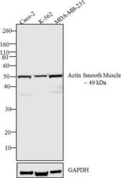
- Western blot analysis was performed on whole cell extracts (30 µg lysate) of Caco-2 (Lane 1), K-562 (Lane 2) and MDA-MB-231 (Lane 3). The blots were probed with Anti-Actin smooth muscle Rabbit Polyclonal Antibody (Product # PA5-16697, 1-3 µg/mL) and detected by chemiluminescence using Goat anti-Rabbit IgG (H+L) Superclonal™ Secondary Antibody, HRP conjugate (Product # A27036, 0.4 µg/mL, 1:2500 dilution). A 49 kDa band corresponding to Actin smooth muscle was observed across the cell lines tested. Known quantity of protein samples were electrophoresed using Novex® NuPAGE® 12 % Bis-Tris gel (Product # NP0342BOX), XCell SureLock™ Electrophoresis System (Product # EI0002) and Novex® Sharp Pre-Stained Protein Standard (Product # LC5800). Resolved proteins were then transferred onto a nitrocellulose membrane with PierceTM Power Blotter System (Product # 22834). The membrane was probed with the relevant primary and secondary Antibody using iBind™ Flex Western Starter Kit (Product # SLF2000S). Chemiluminescent detection was performed using Pierce™ ECL Western Blotting Substrate (Product # 32106).

- Submitted by
- Invitrogen Antibodies (provider)
- Main image
- Experimental details
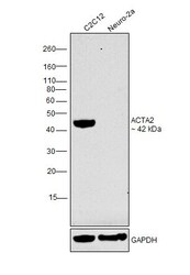
- Western blot was performed using Anti-Alpha-Smooth Muscle Actin Polyclonal Antibody (Product # PA5-16697) and a 42 kDa band corresponding to ACTA2 was observed in C2C12, the positive model for ACTA2 while not detected in Neuro-2a which is reported to be low for ACTA2, relative to C2C12. Membrane enriched extracts (30 µg lysate) of C2C12 (Lane 1) and Neuro-2a (Lane 2) were electrophoresed using NuPAGE™ 10% Bis-Tris Protein Gel (Product # NP0302BOX). Resolved proteins were then transferred onto a nitrocellulose membrane (Product # IB23001) by iBlot® 2 Dry Blotting System (Product # IB21001). The blot was probed with the primary antibody (1µg/ml) and detected by chemiluminescence with Goat anti-Rabbit IgG (H+L), Superclonal™ Recombinant Secondary Antibody, HRP (Product # A27036, 1:4000 dilution) using the iBright FL 1000 (Product # A32752). Chemiluminescent detection was performed using Novex® ECL Chemiluminescent Substrate Reagent Kit (Product # WP20005)..
Supportive validation
- Submitted by
- Invitrogen Antibodies (provider)
- Main image
- Experimental details
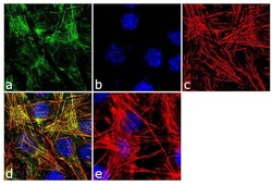
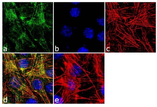
- Immunofluorescent analysis of Actin Smooth Muscle was performed using 70% confluent log phase C2C12 cells. The cells were fixed with 4% paraformaldehyde for 10 minutes, permeabilized with 0.1% Triton™ X-100 for 10 minutes, and blocked with 1% BSA for 1 hour at room temperature. The cells were labeled with Actin Smooth Muscle Rabbit Polyclonal Antibody (Product # PA5-16697) at 2 µg/mL in 0.1% BSA and incubated for 3 hours at room temperature and then labeled with Goat anti-Rabbit IgG (H+L) Superclonal™ Secondary Antibody, Alexa Fluor® 488 conjugate (Product # A27034) a dilution of 1:2000 for 45 minutes at room temperature (Panel a: green). Nuclei (Panel b: blue) were stained with SlowFade® Gold Antifade Mountant with DAPI (Product # S36938). F-actin (Panel c: red) was stained with Alexa Fluor® 555 Rhodamine Phalloidin (Product # R415, 1:300). Panel d represents the merged image showing cytoskeleton localization. Panel e shows the no primary antibody control. The images were captured at 60X magnification.
Supportive validation
- Submitted by
- Invitrogen Antibodies (provider)
- Main image
- Experimental details
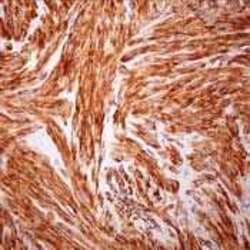
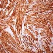
- Formalin-fixed, paraffin-embedded human leiomyoma stained with Actin using peroxidase-conjugate and AEC. Note cytoplasmic staining of tumor cells.
Supportive validation
- Submitted by
- Invitrogen Antibodies (provider)
- Main image
- Experimental details
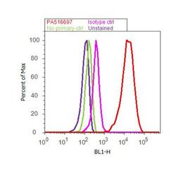
- Flow cytometry analysis of Actin Smooth Muscle was done on C2C12 cells. Cells were fixed with 70% ethanol for 10 minutes, permeabilized with 0.25% Triton™ X-100 for 20 minutes, and blocked with 5% BSA for 30 minutes at room temperature. Cells were labeled with Actin Smooth Muscle Rabbit Polyclonal Antibody (PA5-16697, red histogram) or with rabbit isotype control (pink histogram) at 3-5 ug/million cells in 2.5% BSA. After incubation at room temperature for 2 hours, the cells were labeled with Alexa Fluor® 488 Goat Anti-Rabbit Secondary Antibody (A11008) at a dilution of 1:400 for 30 minutes at room temperature. The representative 10, 000 cells were acquired and analyzed for each sample using an Attune® Acoustic Focusing Cytometer. The purple histogram represents unstained control cells and the green histogram represents no-primary-antibody control.
Supportive validation
- Submitted by
- Invitrogen Antibodies (provider)
- Main image

- Experimental details
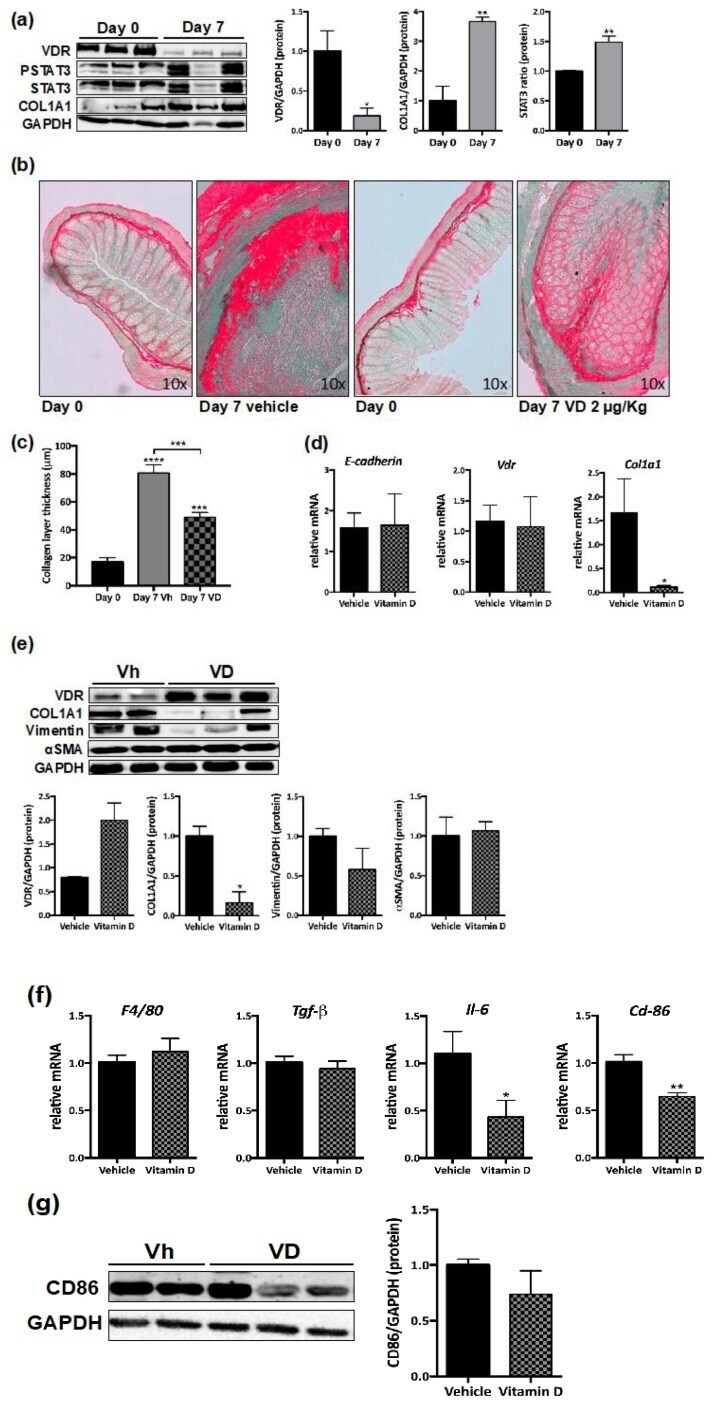
- Figure 4 VD reduces murine intestinal fibrosis. ( a ) Western blots of protein levels in total lysates from intestinal grafts at day 0 (control) ( n = 3) or seven days after transplantation ( n = 3). Graphs show protein expression vs. GAPDH represented as fold induction vs. day 0. Bars in graph represent mean +- s.e.m., and significant differences vs. day 0 are shown by * p < 0.05 or ** p < 0.01 ( b ) Sirius Red staining was performed in paraffin-embedded intestinal tissue at day 0 and in intestinal explants. Representative pictures taken under transmission light. ( c ) Graph shows the collagen layer thickness quantified in intestine and grafts by Image J. Significant differences vs. day 0 or vs. 7 days-vehicle (connecting lines) are shown by *** p < 0.001. ( d ) Graphs show the relative mRNA expression (expressed as fold induction vs. vehicle-treated group) of different genes vs. beta-actin in intestinal explants from mice treated for 7 days with VD 2 mug/kg ( n = 6) or vehicle ( n = 6). ( e ) Western blot images of protein expression from 7 day grafts from mice treated with VD ( n = 3) or vehicle ( n = 2). Graphs represent protein expression vs. GAPDH quantification expressed as fold induction vs. vehicle-treated group. In ( d ) and ( e ) , bars in graph represent mean +- s.e.m. and significant differences vs. the vehicle group are shown by * p < 0.05. ( f ) Graphs show the mRNA expression of different genes vs. beta-Actin (expressed as fold induction vs. vehicle) in 7 day
- Submitted by
- Invitrogen Antibodies (provider)
- Main image
- Experimental details
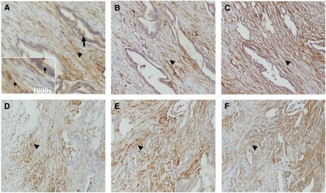
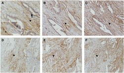
- Figure 2 Representative images of immunohistochemical staining patterns on consecutive tissue sections demonstrating the presence of (A and D) urokinase plasminogen receptor, (B and E) vimentin, and (C and F) alpha-smooth muscle actin in pancreatic adenocarcinoma (x200 magnification). Arrows and arrow heads indicate, respectively, epithelial and stromal cells. The insert in A represents the x1000 magnification of the area with arrow and arrow head. uPAR, vimentin were stained with mouse monoclonal antibodies ATN-615 and V9. Smooth muscle actin was detected by rabbit polyclonal antibodies (PA5-16697).