Explore
 Validate
 Learn
Learn Western blot
 Other assay
Antibody data
- Antibody Data
- Antigen structure
- References [1]
- Comments [0]
- Validations
- Other assay [9]
Submit
Validation data
Reference
Comment
Report error
- Product number
- PA5-69223 - Provider product page

- Provider
- Invitrogen Antibodies
- Product name
- MARCH1 Polyclonal Antibody
- Antibody type
- Polyclonal
- Antigen
- Synthetic peptide
- Description
- This target displays homology in the following species: Cow: 100%; Dog: 100%; Guinea Pig: 100%; Horse: 100%; Human: 100%; Mouse: 100%; Rabbit: 100%; Rat: 100%
- Reactivity
- Human
- Host
- Rabbit
- Isotype
- IgG
- Vial size
- 100 μL
- Concentration
- 0.5 mg/mL
- Storage
- -20°C, Avoid Freeze/Thaw Cycles
Submitted references Elucidating the Antiviral Mechanism of Different MARCH Factors.
Umthong S, Lynch B, Timilsina U, Waxman B, Ivey EB, Stavrou S
mBio 2021 Mar 2;12(2)
mBio 2021 Mar 2;12(2)
No comments: Submit comment
Supportive validation
- Submitted by
- Invitrogen Antibodies (provider)
- Main image
- Experimental details
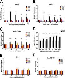
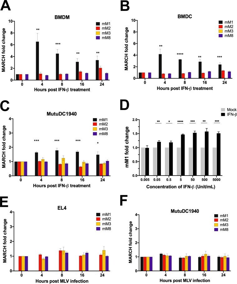
- FIG 1 Expression levels of mouse March1 , 2 , 3 , and 8 in the presence of IFN-beta and MLV infection. (A to C) Fold expression changes of mouse March1 , 2 , 3 , and 8 ( mM1 , 2 , 3 , and 8 ) relative to untreated cells and normalized to GAPDH in (A) bone marrow-derived macrophages (BMDMs), (B) bone marrow-derived dendritic cells (BMDCs), and (C) MutuDC1940 cells at 0, 4, 8, 16, and 24 h after treatment with 500 units/ml of mouse IFN-beta. Purity of BMDMs and BMDCs was determined by flow cytometry staining with anti-CD11b and anti-CD11c, respectively, as shown in Fig. S1D and E in the supplemental material. (D) Fold expression changes of mM1 relative to untreated cells and normalized to GAPDH after treatment of MutuDC1940 cells with different concentrations of IFN-beta (0.005 to 5,000 units/ml) for 4 h. (E and F) Fold expression changes of mM1, 2, 3, and 8 relative to uninfected cells and normalized to GAPDH in (E) mouse T lymphocyte cell line EL4 and (F) MutuDC1940 cells at 4, 8, 16, and 24 h postinfection with MLV (5 MOI). Data shown represent averages of results from n = 3 independent experiments. All results are presented as means +- standard error of the mean (SEM). Statistical analysis performed using unpaired t test. *, P
- Submitted by
- Invitrogen Antibodies (provider)
- Main image

- Experimental details
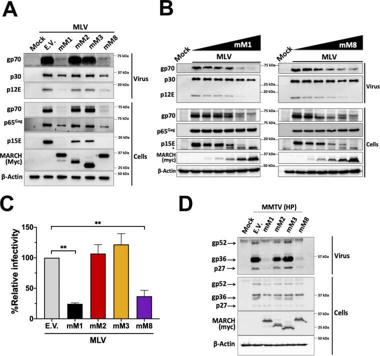
- FIG 2 Mouse March1 and mouse March8 block retrovirus infection by targeting the retroviral envelope glycoproteins. (A) Mouse March1 (mM1) and mM8 block the incorporation of the MLV envelope glycoproteins (p15E and gp70) in nascent virions by targeting them for degradation. (B) mM1 and mM8 target the MLV envelope glycoproteins for degradation in a dose-dependent manner. For both panels A and B, 293T cells were cotransfected with an MLV molecular clone and the indicated mouse March constructs. At 48 h posttransfection, cells and released virus in the culture medium were harvested, and the indicated proteins were analyzed by immunoblotting using anti-MLV p30 (detects both p30 and p65 Gag ), anti-MLV gp70, anti-MLV p15E/p12E, anti-myc (for detection of mM1, 2, 3, and 8), and anti-beta-actin antibodies. (C) Virus produced in the presence of mM1 and mM8 has decreased infectivity. NIH 3T3 cells were infected with equal amounts of 293T-derived MLV-luciferase reporter virus produced in the presence of mM1, 2, 3, or 8 or empty vector (E.V.). Cells were harvested 24 h postinfection, and luciferase levels were measured. The percentage (%) of relative infectivity was determined with respect to virus produced in the presence of E.V. All results are presented as means +- SD. Statistical significance was determined by one-way ANOVA. **, P < 0.01. (D) mM1 and mM8 target MMTV gp36 for degradation. 293T cells were cotransfected with an infectious genetically engineered MMTV hybrid provirus (HP)
- Submitted by
- Invitrogen Antibodies (provider)
- Main image
- Experimental details
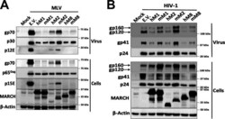
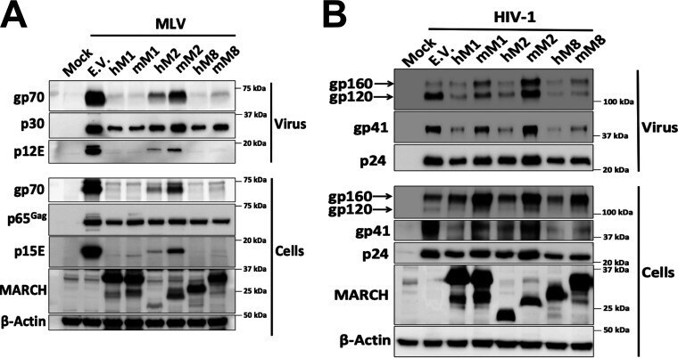
- FIG 3 Mouse March and human MARCH proteins differ in their ability to target retroviral envelope glycoproteins. (A) MLV envelope glycoproteins are targeted for degradation by mouse March (mM1) and mM8 and human MARCH (hM1), hM2, and hM8. (B) HIV-1 envelope glycoproteins are targeted for degradation by mM8 and hM1, 2, and 8. For both panels A and B, 293T cells were cotransfected with (A) MLV or (B) HIV-1 infectious clone and hmM1 , 2 , or 8 . At 48 h posttransfection, cells and released virus in the culture medium were harvested, and the indicated proteins were analyzed by immunoblotting using anti-MLV p30 (detects both p30 and p65 Gag ), anti-MLV gp70, anti-MLV p15E/p12E, anti-HIV-1 envelope (detects both gp120 and gp160), anti-gp41, anti-p24, anti-myc (for detection of mM1, 2, and 8), anti-MARCH1, 2, 8 and anti-beta-actin antibodies. Results are shown for n = 3 independent experiments. Representative immunoblotting results are shown for panels A and B.
- Submitted by
- Invitrogen Antibodies (provider)
- Main image
- Experimental details
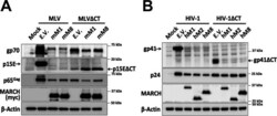
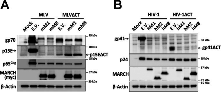
- FIG 5 The MLV envelope glycoprotein cytoplasmic tail is critical for MARCH-mediated restriction, but the HIV envelope cytoplasmic tail is dispensable. 293T cells were cotransfected in panel A with wild-type MLV or MLV with the cytosolic tail deleted (MLVDeltaCT) along with either mouse March1 (mM1), mM2, or mM8 and in panel B with wild-type HIV or HIV with the cytosolic tail deleted (HIVDeltaCT) along with either human MARCH1 (hM1), hM2, or hM8. For panels A and B, at 48 h posttransfection cells were harvested and lysates were analyzed by immunoblotting using anti-MLV p30 (detects p65 Gag ), anti-MLV gp70, anti-MLV p15E, anti-myc (for detection of human and mouse MARCH proteins), anti-gp41, anti-p24, and anti-beta-actin antibodies. Results are shown for n = 3 independent experiments. Representative immunoblotting results are shown for panels A and B.
- Submitted by
- Invitrogen Antibodies (provider)
- Main image
- Experimental details
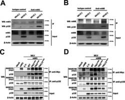
- FIG 6 Mouse March1 and mouse March8 physically interact with MLV p15E. (A and B) MutuDC1940 cells, which express endogenous levels of both mouse March1 (mM1) and mM8, were infected with MLV (10 MOI). Cells were harvested 72 h postinfection, and lysates were immunoprecipitated with (A and B) an isotype control, (A) anti-MARCH1, and (B) anti-MARCH8 and analyzed in Western blots (WB) with anti-p15E, anti-MARCH1, anti-MARCH8, and anti-beta-actin. (C and D) 293T cells were cotransfected with an MLV infectious clone and either (C) mouse March1 ( mM1 ) or (D) mM8 and their TM mutants used in Fig. 4 . Cells were harvested 48 h posttransfection, and lysates were immunoprecipitated with anti-myc (mM1 and mM8) and anti-p15E followed by Western blot analyses probing with anti-p15E, anti-myc (mM1 and mM8), and anti-beta-actin antibodies. Results are shown for n = 3 independent experiments. Representative immunoblotting results are shown for panels A through D.

- Submitted by
- Invitrogen Antibodies (provider)
- Main image
- Experimental details
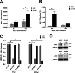
- FIG 8 Endogenous mouse March1 and mouse March8 restrict MLV infection. (A and B) Bone marrow-derived dendritic cells (BMDCs) were transfected with the indicated siRNAs and infected with MLV (0.1 MOI). Cells were harvested 6 and 24 h postinfection, and MLV DNA levels were examined by RT-qPCR. (C and D) Knockdown verification of mouse March1 (mM1) and mM8 from siRNA-transfected BMDCs relative to siControl (siCtl) in panels A and B by either (C) RT-PCR or (D) immunoblotting. Data shown represent averages of results from n = 5 independent experiments. Results for panels A through C are presented as means +- standard errors of the means (SEM). Representative immunoblotting results are shown for panel D. ImageJ (NIH) was used for quantitation of the mM1 and mM8 protein levels. Statistical analysis performed using one-sample t test and Wilcoxon signed-rank test and unpaired t test. *, P

- Submitted by
- Invitrogen Antibodies (provider)
- Main image
- Experimental details
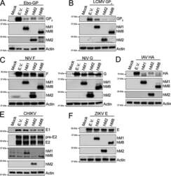
- FIG 9 MARCH proteins target envelope glycoproteins from a diverse number of viral families. (A) Ebola virus (EBOV) mature envelope glycoprotein Gp 2 , (B) lymphocytic choriomeningitis virus (LCMV) mature envelope glycoprotein GP 2 , (C) Nipah virus (NiV) fusion glycoprotein (F) (left) and NiV receptor binding glycoprotein (G) (right), (D) influenza A virus (IAV) hemagglutinin (HA), (E) Chikungunya virus (CHIKV) E1 and E2 protein, and (F) Zika virus (ZIKV) E levels in the presence of human MARCH1 (hM1), MARCH2 (hM2), MARCH8 (hM8), and empty vector (E.V). 293T cells were cotransfected with either (A) EBOV glycoprotein (Gp), (B) LCMV Gp, (C) NiV F or NiV G, (D) IAV HA, (E) a plasmid expressing the CHIKV structural proteins, or (F) ZIKV E in combination with either hM1 , 2 , 8 , or E.V. At 24 h posttransfection, cells were lysed and analyzed by Western blotting using anti-V5 (for EBOV GP 2 and IAV HA detection), anti-FLAG (for LCMV GP 2 detection), anti-AU1 (for NiV G detection), anti-HA (for NiV F detection), anti-flavivirus E antibody (4G2, for ZIKV E detection), anti-myc (CHIKV E1 detection), anti-E2 (CHIKV E2 detection), anti-MARCH1, anti-MARCH2, anti-MARCH8, and anti-beta-actin antibodies. Shown are the results of a single experiment (representative of three independent experiments).

- Submitted by
- Invitrogen Antibodies (provider)
- Main image
- Experimental details
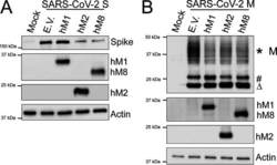
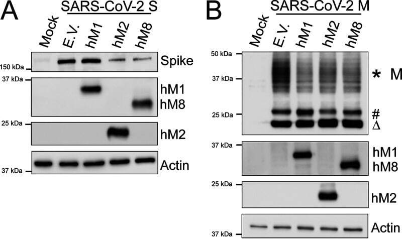
- FIG 10 MARCH proteins potently restrict the SARS-CoV-2 Spike (S) and Membrane (M) envelope glycoproteins. (A) SARS-CoV-2 Spike (S) and (B) SARS-CoV-2 Membrane (M) cellular levels in the presence of human MARCH1 (hM1), MARCH2 (hM2), MARCH8 (hM8), and empty vector (E.V.). 293T cells were cotransfected with SARS-CoV-2 S (A) or M (B) and either hM1 , 2 , 8 , or E.V. At 24 h posttransfection, cells were lysed and analyzed by Western blotting using anti-SARS-CoV-2 S, anti-V5 (for SARS-CoV-2 M detection), anti-MARCH1, anti-MARCH2, anti-MARCH8, and anti-beta-actin antibodies. The *, #, and Delta symbols represent different glycosylated forms of SARS-CoV-2 M. Shown are the results of a single experiment (representative of three independent experiments).
- Submitted by
- Invitrogen Antibodies (provider)
- Main image

- Experimental details
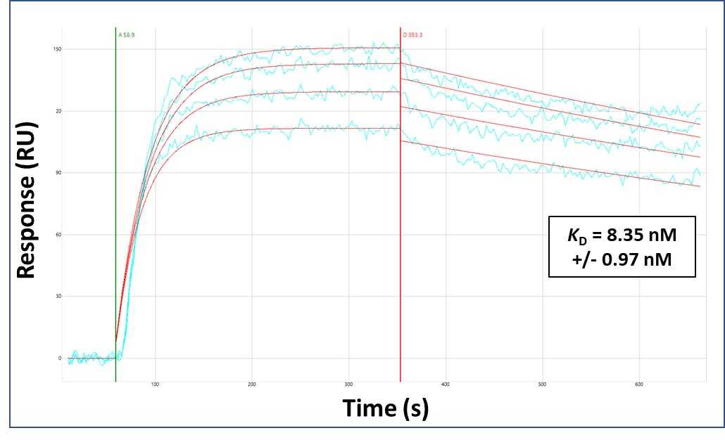
- Surface Plasmon Resonance of MARCH1 polyclonal antibody (Product # PA5-69223). Purified polyclonal antibodies were immobilized on a Protein A/G coated Carterra LSA sensor chip at concentrations of 5, and 50 µg/mL in duplicate. Antibodies on the surface were exposed to interaction with peptides sequentially via microfluidic controlled flow at 333 nm peptide concentration for kinetic characterization of the binders for affinity and specificity, followed by curve fitting using the Kinetics software. Kd determinations for both concentrations were averaged and results and standard deviation are shown.