 Explore
 Validate
 Learn
Learn Western blot
Antibody data
- Antibody Data
- Antigen structure
- References [0]
- Comments [0]
- Validations
- Western blot [2]
- Immunocytochemistry [1]
- Immunohistochemistry [1]
Submit
Validation data
Reference
Comment
Report error
- Product number
- PA5-47894 - Provider product page

- Provider
- Invitrogen Antibodies
- Product name
- FUCA1 Polyclonal Antibody
- Antibody type
- Polyclonal
- Antigen
- Recombinant full-length protein
- Description
- Reconstitute in sterile PBS to a final concentration of 0.2 mg/mL.
- Reactivity
- Human
- Host
- Sheep
- Isotype
- IgG
- Vial size
- 100 µg
- Concentration
- 0.2 mg/mL
- Storage
- -20° C, Avoid Freeze/Thaw Cycles
No comments: Submit comment
Supportive validation
- Submitted by
- Invitrogen Antibodies (provider)
- Main image

- Experimental details
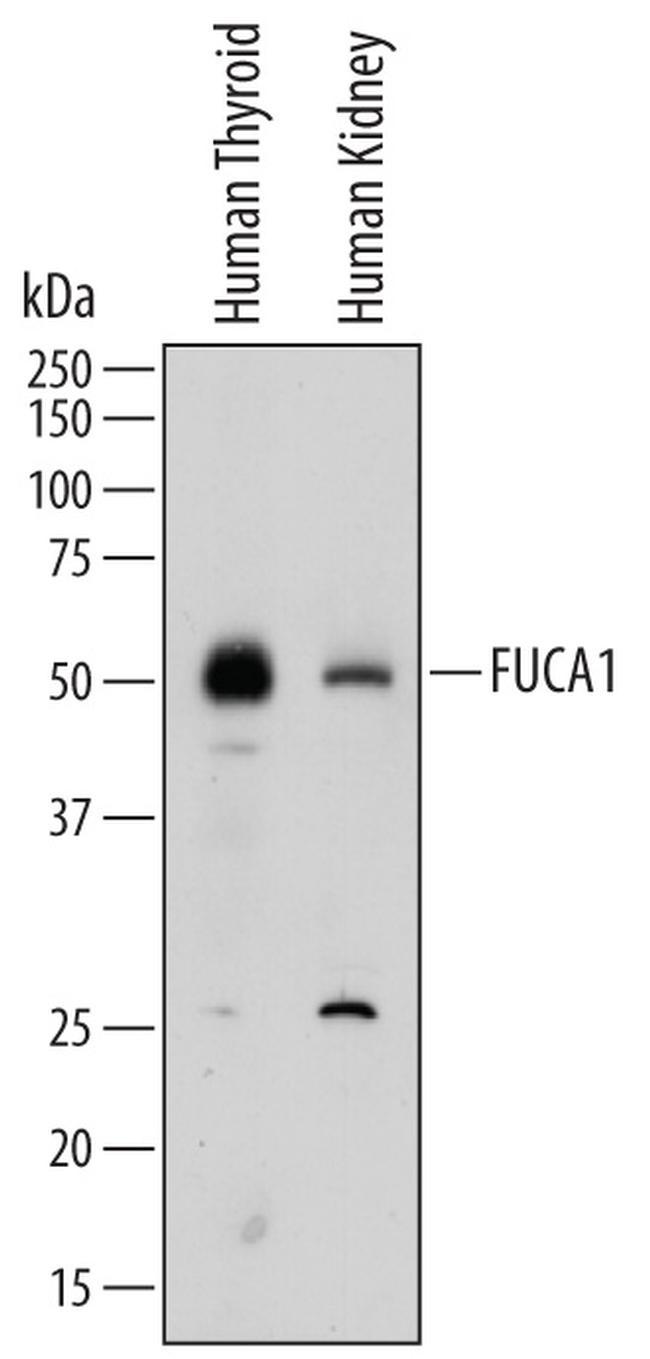
- Western blot analysis from lysates of human thyroid tissue and human kidney tissue. PVDF membrane was probed with 1 µg/mL of Sheep Anti-human Tissue a-L-Fucosidase/FUCA1 Antigen Affinity-purified Polyclonal Antibody (Product # PA5-47894) followed by HRP-conjugated Anti-Sheep IgG Secondary Antibody. A specific band was detected for Tissue a-L-Fucosidase/FUCA1 at approximately 50 kDa (as indicated). This experiment was conducted under reducing conditions.
- Submitted by
- Invitrogen Antibodies (provider)
- Main image
- Experimental details
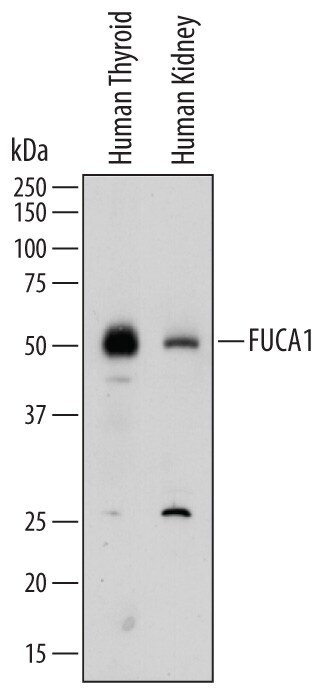
- Western blot analysis of FUCA1 in human thyroid tissue and human kidney tissue. Samples were incubated in FUCA1 polyclonal antibody (Product # PA5-47894) using a dilution of 1 µg/mL followed by a HRP-conjugated Anti-Sheep IgG secondary antibody. A specific band was detected for Tissue α‚L‚Fucosidase/FUCA1 at approximately 50 kDa (as indicated). This experiment was conducted under reducing conditions.
Supportive validation
- Submitted by
- Invitrogen Antibodies (provider)
- Main image

- Experimental details
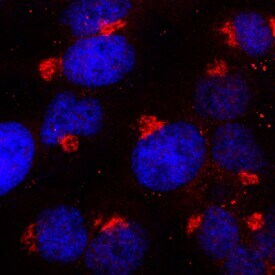
- Immunocytochemistry analysis of FUCA1 in immersion fixed HeLa human cervical epithelial carcinoma cell line. Samples were incubated in FUCA1 polyclonal antibody (Product # PA5-47894) using a dilution of 15 µg/mL for 3 hours at room temperature followed by NorthernLights™ 557-conjugated Anti-Sheep IgG Secondary Antibody (red) and counterstained with DAPI (blue). Specific staining was localized to lysosomes.
Supportive validation
- Submitted by
- Invitrogen Antibodies (provider)
- Main image
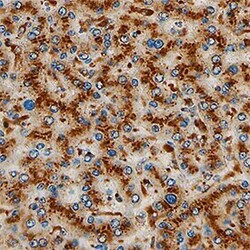
- Experimental details
- Immunohistochemical analysis of FUCA1 in immersion fixed paraffin-embedded sections of human liver. Samples were incubated with FUCA1 polyclonal antibody (Product # PA5-47894) using a dilution of 5 µg/mL for 1 hour at room temperature followed by Anti-Sheep IgG VisUCyte™ HRP Polymer Antibody. Tissue was stained using DAB (brown) and counterstained with hematoxylin (blue). Specific staining was localized to lysosomes in hepatocytes.
